. 2010 Sep;30(17):4175-87.
doi: 10.1128/MCB.00274-10. Epub 2010 Jun 28.

Sortilin facilitates signaling of ciliary neurotrophic factor and related helical type 1 cytokines targeting the gp130/leukemia inhibitory factor receptor beta heterodimer

Affiliations

Sortilin facilitates signaling of ciliary neurotrophic factor and related helical type 1 cytokines targeting the gp130/leukemia inhibitory factor receptor beta heterodimer

Jakob Vejby Larsen et al. Mol Cell Biol. 2010 Sep.

Abstract

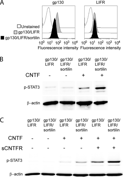
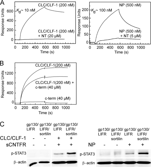
Sortilin is a member of the Vps10p domain family of neuropeptide and neurotrophin binding neuronal receptors. The family members interact with and partly share a variety of ligands and partake in intracellular sorting and protein transport as well as in transmembrane signal transduction. Thus, sortilin mediates the transport of both neurotensin and nerve growth factor and interacts with their respective receptors to facilitate ligand-induced signaling. Here we report that ciliary neurotrophic factor (CNTF), and related ligands targeting the established CNTF receptor alpha, binds to sortilin with high affinity. We find that sortilin may have at least two functions: one is to provide rapid endocytosis and the removal of CNTF, something which is not provided by CNTF receptor alpha, and the other is to facilitate CNTF signaling through the gp130/leukemia inhibitory factor (LIF) receptor beta heterodimeric complex. Interestingly, the latter function is independent of both the CNTF receptor alpha and ligand binding to sortilin but appears to implicate a direct interaction with LIF receptor beta. Thus, sortilin facilitates the signaling of all helical type 1 cytokines, which engage the gp130/LIF receptor beta complex.


Figures

FIG. 1.
FIG. 1.
Sortilin binds and mediates cellular uptake of CNTF. Purified s-sortilin and s-sortilin containing an uncleavable propeptide (s-prosortilin) were immobilized on Biacore chips. CNTF binding was then analyzed by SPR. (A) Concentration dependence of binding. CNTF was applied at the given concentrations, and the indicated Kd values were calculated on the basis of the collected sum of data. (B) Binding of CNTF in the absence or presence of a 50- to 100-fold excess of NT. (C) Inhibition of CNTF binding by RAP. Sortilin was saturated with RAP (at 10 μM) prior to the injection of a mixture of RAP (10 μM) and CNTF (200 nM). For comparison, the response obtained with CNTF alone has been moved to the level of RAP saturation. (D) CNTF binding to similar concentrations of wt sortilin and mutant prosortilin. (E) Sortilin binding to sCNTFRα and a preformed complex of sCNTFRα and CNTF. Soluble CNTFRα was immobilized on a Biacore chip and then saturated with CNTF (at 1 μM) prior to the injection of a mixture of CNTF (1 μM) and sortilin (200 nM). The binding of sortilin (200 nM) alone to CNTFRα is shown. (F to J) HEK293 cells, untransfected (F) or stably transfected with either sortilin (G and H), mutant sortilin containing disrupted internalization signals (I), or uncleavable prosortilin (J), were incubated (37°C) with 50 nM CNTF (F, G, I, and J) or with 50 nM CNTF plus 20 μM NT (H). After 45 min the cells were fixed, incubated with rabbit anti-CNTF and mouse antisortilin, and finally stained by using Alexa 488-conjugated goat anti-rabbit Ig (green) and Alexa 633-conjugated goat anti-mouse Ig (red). Nuclei were stained with DAPI (blue). CNTF staining alone is shown in the insets.
FIG. 2.
FIG. 2.
Bound CNTF is rapidly internalized by HEK293 transfectants. Untransfected HEK293 cells (A and B) and stably transfected HEK293 cells expressing either mutant sortilin containing disrupted internalization motifs (C and D) or wt sortilin (E and F) were incubated (4°C) with 50 nM CNTF for 2 h prior to washing and reincubation in warm (37°C) medium. After 10 min the cells were fixed and stained using rabbit anti-CNTF and mouse antisortilin as primary antibodies and Alexa 488-conjugated goat anti-rabbit Ig (green) and Alexa 633-conjugated goat anti-mouse Ig (red) as secondary antibodies. Nuclei were stained with DAPI (blue). Double staining is shown in the insets.
FIG. 3.
FIG. 3.
C-terminally truncated CNTF does not bind sortilin. Purified s-sortilin was immobilized on a Biacore chip. The binding of CNTF-tr and full-length CNTF was determined by SPR analysis. (A) Binding of 500 nM CNTF-tr and wt CNTF. (B) Binding of wt CNTF in the absence and presence of a 20- to 40-fold excess of a 13-residue peptide constituting the C terminus of CNTF. (C to E) HEK293 cells stably transfected with sortilin were incubated (37°C) with 50 nM CNTF-tr (C) and 50 nM wt CNTF with or without 50 μM C-terminal peptide (D and E). After 45 min the cells were fixed, incubated with rabbit anti-CNTF, and finally stained by using Alexa 488-conjugated goat anti-rabbit Ig (green). Nuclei were stained with DAPI (blue). (F and G) Binding of CNTF (F) and CNTF-tr (G) to immobilized CNTFRα. The CNTF constructs were applied at the given concentrations, and the indicated Kd values were calculated on the basis of the collected sum of data.
FIG. 4.
FIG. 4.
Sortilin enhances the CNTF-induced phosphorylation of STAT3 in TF-1 cells. (A) FACS analysis showing the expression of gp130 and LIFRβ on wt TF-1 cells and on sortilin-transfected TF-1 cells (TF-1/sort). The right panel shows a Western blot performed with anti-CNTFRα Ig on lysates of untransfected TF-1 cells and (as control) BA/F3 transfectants expressing CNTFRα. (B) TF-1 cells, wt or transfected with sortilin, were exposed to 40 nM CNTF for 15 min. The cells were subsequently lysed, and the levels of phosphorylated STAT3 (p-STAT3) and β-actin (control) were determined by Western blotting. (C) Histogram showing the level of phospho-STAT3 in unstimulated or stimulated (40 nM CNTF) wt TF-1 and sortilin-transfected TF-1 cells. Levels were determined by the quantification of Western blots, and values are shown relative to the level observed for unstimulated TF-1 cells (set to 1). Results of eight separate experiments are shown. (D) Western blot performed with antisortilin Ig on lysates of untransfected TF-1 cells or cells stably transfected with either wt sortilin or mutant sortilin without the cytoplasmic domain (TF-1/sort-cd). (E) TF-1 cells, wt, sortilin transfected, or transfectants expressing mutant sortilin lacking the cytoplasmic domain, were incubated with 40 nM CNTF. After 15 min the cells were lysed, and the levels of phosphorylated STAT3 and β-actin were determined.
FIG. 5.
FIG. 5.
CNTF signaling in wt and transfected BA/F3 cells. (A) FACS analysis of receptors (indicated) on the surface membrane of BA/F3 transfectants expressing gp130 and LIFRβ alone or in combination with sortilin (filled gray and black curves, respectively). (B) Western blot showing sortilin in lysates of BA/F3 cells expressing gp130 and LIFRβ before and after their transfection with sortilin and immunofluorescence of the same cells by using monoclonal antisortilin Ig and Alexa 488-conjugated goat anti-mouse antibody (green) as primary and secondary antibodies. Nuclei are indicated by DAPI staining (blue). (C) BA/F3 cells expressing the indicated combinations of gp130, LIFRβ, sortilin, and CNTFRα were incubated in the absence (lanes 1 to 7) or presence (lanes 8 to 14) of 40 nM CNTF for 15 min. The cells were then lysed, and their contents of phosphorylated STAT3 and β-actin were determined by Western blotting. (D) Histogram showing the quantification of phosphorylated STAT3 (determined by Western blots) in BA/F3-[gp130/LIFRβ] and BA/F3-[gp130/LIFRβ/sortilin] cells relative to the level obtained in unstimulated BA/F3-[gp130/LIFRβ] cells. Results are presented as mean values (±SD) from 22 separate experiments. (E) The samples from lanes 6, 7, 13, and 14 in C were also probed for MAP kinase activation using anti-phosphorylated MAP kinase (anti-p-MAP kinase) antibodies. (F) Time course of 40 nM CNTF-induced STAT3 phosphorylation in BA/F3 cells transfected with gp130, LIFRβ, and sortilin. The cells were stimulated with CNTF for the times indicated and then lysed. The levels of STAT3 and β-actin in the cell lysates were then determined by Western blotting.
FIG. 6.
FIG. 6.
Sortilin enhances the CNTF-induced phosphorylation of STAT3 without upregulating gp130 and LIFRβ cell surface levels. (A) Surface expression of gp130 and LIFRβ in BA/F3-[gp130/LIFRβ] (gray) and BA/F3-[gp130/LIFRβ/sortilin] (black) cells. A comparatively low level of gp130 was seen in the sortilin transfectants. (B) Phospho-STAT3 levels upon stimulation of the same cells with 40 nM CNTF (15 min at 37°C). Stimulation was performed in parallel with FACS analysis. (C) BA/F3 transfectants expressing the indicated receptor combinations were incubated with or without 40 nM CNTF and 4 nM sCNTFRα for 15 min at 37°C. The cells were then lysed, and the amounts of phospho-STAT3 and β-actin contained in the lysates were determined by Western blotting.
FIG. 7.
FIG. 7.
Influence of sortilin and sCNTFRα on CNTF-mediated BA/F3 cell proliferation. BA/F3 transfectants expressing the given combinations of receptors were incubated in 96-well plates (20 × 104 cells per well) containing various concentrations of CNTF alone or in combination with 4 nM sCNTFRα (inset). After 72 h, [3H]thymidine was added to each well, and after an additional 24 h of incubation, the cells were harvested, and the incorporated radioactivity was determined with a Beckman β-counter. Each point represents the mean (±SD) of data from three separate experiments. The inset graph shows data from a representative experiment, where each point is the mean (±SD) of data from triplicate samples.
FIG. 8.
FIG. 8.
Sortilin binds and enhances the signaling of CLC/CLF-1 and neuropoietin. (A) SPR analysis of CLC/CLF-1 (left) and neuropoietin (NP) (right) binding to immobilized s-sortilin. The responses obtained in both the absence and presence of NT-mediated inhibition are shown, and the estimated Kd values are indicated. (B) Binding of CLC/CLF-1 in the absence or presence of a 200-fold excess of the 13-residue peptide constituting the C terminus of CNTF. (C) STAT3 phosphorylation induced by CLC/CLF-1 (left) and neuropoietin (right). BA/F3 transfectants expressing the indicated combinations of receptors were stimulated for 15 min at 37°C with CLC/CLF-1 (40 nM) plus sCNTFRα (16 nM) or neuropoietin (1 nM) prior to Western blotting of lysed cells.
FIG. 9.
FIG. 9.
Sortilin enhances CNTF signaling independent of ligand binding. (A) BA/F3 cells expressing gp130 and LIFRβ alone or in combination with sortilin were incubated for 15 min at 37°C with or without 40 nM CNTF. The incubations were carried out in the absence or presence of 40 μM NT, and the resulting phosphorylation of STAT3 was determined by Western blotting of cell lysates. (B) TF-1 cells, wt or transfected with sortilin, were incubated for 15 min at 37°C with or without 40 nM CNTF in the absence or presence of 9 μM RAP, 40 μM NT, and 50 μM sortilin GST-propeptide (pro) prior to Western blot analysis of lysed cells.
FIG. 10.
FIG. 10.
CNTF-tr induces phosphorylation of STAT3 in TF-1 and BA/F3 cells. (A) TF-1 cells, wt or sortilin transfected, were incubated with 40 nM CNTF-tr and full-length CNTF. After 15 min the cells were lysed, and the levels of phosphorylated STAT3 and β-actin were determined. (B) BA/F3 cells expressing gp130 and LIFRβ in combination with sortilin or CNTFRα were incubated for 15 min at 37°C with or without 40 nM CNTF-tr and full-length CNTF prior to Western blot analysis of lysed cells. (C) BA/F3 cells transfected with gp130, LIFRβ, and sortilin were incubated with or without CNTF in the absence or presence of 50 μM C-term and then lysed. The levels of STAT3 and β-actin in the cell lysates were finally determined by Western blotting.
FIG. 11.
FIG. 11.
Sortilin enhances signaling of CT-1, LIF, and OSM but not IL-6. (A) TF-1 cells, wt and transfected with sortilin, were incubated with or without 1 nM CT-1, LIF, and OSM. After 15 min the cells were then lysed, and their contents of phosphorylated STAT3 and β-actin were determined by Western blotting. (B) BA/F3 cells expressing gp130 and LIFRβ alone or in combination with sortilin were incubated for 15 min at 37°C with or without 0.1 nM CT-1, LIF, and OSM prior to Western blotting of lysed cells. (C) TF-1 cells, wt or sortilin transfected, were incubated with or without 1 nM or 0.1 nM hIL-6 for 15 min followed by Western blotting of lysed cells. (D) BA/F3 cells expressing gp130 alone or in combination with sortilin were incubated with or without 0.01 nM or 0.001 nM hIL-6 for 15 min. Cell lysates were Western blotted and probed with antibodies against phosphorylated STAT3 and β-actin.
FIG. 12.
FIG. 12.
Interaction and colocalization of LIFRβ and sortilin. (A) Purified s-sortilin was immobilized on a Biacore chip, and the binding of sLIFRβ (200 nM) and sgp130-Fc (200 nM) was then analyzed by SPR. The estimated Kd values are indicated. (B) HEK293 cells that express endogenous LIFRβ and sortilin were incubated with or without 40 nM CNTF for 15 min prior to subcellular fractionation. Fractions were Western blotted and probed with antibodies against LIFRβ, sortilin, flotillin-1, and phosphorylated STAT3. (C) HEK293 cells, untransfected or stably transfected with either sortilin or mutant sortilin containing disrupted internalization signals, were fixed and incubated with either the combination of anti-LIFRβ and antisortilin antibodies or the combination of anti-gp130 and antisortilin antibodies overnight. The next day, the cells were labeled with the commercially available Duolink kit according to the manufacturer's instructions. Individual protein interactions are shown in red, and nuclei are indicated by DAPI staining (blue).

Similar articles

Cited by

References

    1. Aasland, D., B. Oppmann, J. Grotzinger, S. Rose-John, and K. J. Kallen. 2002. The upper cytokine-binding module and the Ig-like domain of the leukaemia inhibitory factor (LIF) receptor are sufficient for a functional LIF receptor complex. J. Mol. Biol. 315:637-646. - PubMed
    1. Adler, R., K. B. Landa, M. Manthorpe, and S. Varon. 1979. Cholinergic neuronotrophic factors: intraocular distribution of trophic activity for ciliary neurons. Science 204:1434-1436. - PubMed
    1. Alexander, W. S., S. Rakar, L. Robb, A. Farley, T. A. Willson, J. G. Zhang, L. Hartley, Y. Kikuchi, T. Kojima, H. Nomura, M. Hasegawa, M. Maeda, L. Fabri, K. Jachno, A. Nash, D. Metcalf, N. A. Nicola, and D. J. Hilton. 1999. Suckling defect in mice lacking the soluble haemopoietin receptor NR6. Curr. Biol. 9:605-608. - PubMed
    1. Buk, D. M., M. Waibel, C. Braig, A. S. Martens, P. C. Heinrich, and L. Graeve. 2004. Polarity and lipid raft association of the components of the ciliary neurotrophic factor receptor complex in Madin-Darby canine kidney cells. J. Cell Sci. 117:2063-2075. - PubMed
    1. Chang, Y., G. Tesco, W. J. Jeong, L. Lindsley, E. A. Eckman, C. B. Eckman, R. E. Tanzi, and S. Y. Guenette. 2003. Generation of the beta-amyloid peptide and the amyloid precursor protein C-terminal fragment gamma are potentiated by FE65L1. J. Biol. Chem. 278:51100-51107. - PubMed

Publication types

LinkOut - more resources