. 2010 Oct 7;116(14):2570-8.
doi: 10.1182/blood-2010-02-268136. Epub 2010 Jun 28.

A novel histidine tyrosine phosphatase, TULA-2, associates with Syk and negatively regulates GPVI signaling in platelets

Affiliations

A novel histidine tyrosine phosphatase, TULA-2, associates with Syk and negatively regulates GPVI signaling in platelets

Dafydd H Thomas et al. Blood. .

Abstract

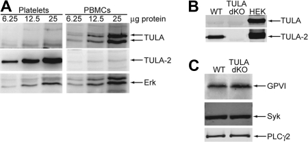
T-cell ubiquitin ligand-2 (TULA-2) is a recently discovered histidine tyrosine phosphatase thought to be ubiquitously expressed. In this work, we have investigated whether TULA-2 has a key role in platelet glycoprotein VI (GPVI) signaling. This study indicates that TULA-2 is expressed in human and murine platelets and is able to associate with Syk and dephosphorylate it. Ablation of TULA-2 resulted in hyperphosphorylation of Syk and its downstream effector phospholipase C-γ2 as well as enhanced GPVI-mediated platelet functional responses. In addition, shorter bleeding times and a prothrombotic phenotype were observed in mice lacking TULA-2. We therefore propose that TULA-2 is the primary tyrosine phosphatase mediating the dephosphorylation of Syk and thus functions as a negative regulator of GPVI signaling in platelets.


Figures

Figure 1
Figure 1
Expression of TULA family members in human and murine platelets and expression of GPVI signaling proteins in murine platelets. (A) Increasing amounts of human platelet and PBMC protein were blotted and probed for TULA family members or the loading control Erk. (B) TULA family protein expression is compared in wild-type and TULA-dKO murine platelets with HEK293T cells overexpressing each member as a positive control. (C) Relative expression of GPVI, Syk, and PLC-γ2 is compared between wild-type and TULA-dKO murine platelets.
Figure 2
Figure 2
TULA-2 dephosphorylation of Syk and association with Syk. (A) Active TULA-2 was purified from Sf9 cells, incubated with phosphorylated Syk for the indicated times, analyzed by Western blotting, and probed for phosphotyrosine, pY525/6 Syk, and total Syk. (B) NP-40 lysed lysates from convulxin-treated and untreated human platelets were precleared and incubated with GST-TULA-2 (H380A) or GST. Pulldowns were analyzed by Western blotting and probed for Syk and TULA-2. (C) NP-40 with 0.1% SDS-lysed lysates from convulxin-treated and untreated human platelets were precleared and incubated with rabbit IgG or anti–TULA-2 antibody. Immunoprecipitates were analyzed by Western blotting and probed for Syk and TULA-2. All blots are representative of 3 experiments.
Figure 3
Figure 3
Syk phosphorylation in wild-type and TULA-dKO murine platelets. (Ai) Isolated wild-type and TULA-dKO platelets were stimulated with 200 ng/mL convulxin for 0, 30, 60 120, and 240 seconds, the protein precipitated, and pY519/20 Syk and total Erk were probed. (Aii) Mean relative Syk phosphorylation of 3 experiments for covulxin in wild-type (■) and TULA-dKO (□) platelets was plotted against time and a 2-way analysis of variance performed (P < .001, WT vs dKO). (Bi) Same as subpanel Ai, except that platelets were stimulated with 1 μg/mL CRP and total Syk was used as a loading control. (Bii) Same as subpanel Aii, except that CRP was used as agonist (P < .001, WT vs dKO). (Ci) Same as subpanel Bi, except that platelets were stimulated with 50 μg/mL collagen. (Di) Isolated wild-type and TULA-dKO platelets were stimulated for 2 minutes with 50, 100, 200, 400, or 800 ng/mL convulxin, processed for Western blotting, and probed for pY519/20 Syk and total Syk. (Dii) Same as subpanel Aii, except that Syk phosphorylation was plotted as a function of convulxin and curves were fitted using a nonlinear regression (P < .001, WT vs dKO). (Ei) Same as subpanel Di, except that platelets were stimulated with 0.5, 1, 2, 5, or 10 μg/mL CRP. (Eii) Same as subpanel Dii, except that CRP was used as agonist (P < .001, WT vs dKO). (F) Same as subpanel Di, except that platelets were stimulated for 1 minute with 25, 50, or 100 μg/mL collagen. (G) Isolated wild-type and TULA dKO platelets were left untreated or treated with 200 ng/mL convulxin for 4 minutes. Kinase activity of the resulting immunoprecipitates was then measured and plotted as function of time. Shown is a representative plot from 5 independent experiments.
Figure 4
Figure 4
PLC-γ2 phosphorylation in wild-type and TULA-dKO murine platelets. (A) Isolated wild-type and TULA-dKO platelets were stimulated with 200 ng/mL convulxin for 0, 30, 60, 120, and 240 seconds, the protein precipitated, analyzed by Western blotting, and pY759 PLC-γ2 and total PLC-γ2 probed. (B) Same as panel A, except that platelets were stimulated with 50 μg/mL collagen. (C) Isolated wild-type and TULA-dKO platelets were stimulated for 2 minutes with 50, 100, 200, 400, or 800 ng/mL convulxin, processed for Western blotting, and probed for pY759 PLC-γ2 and total PLC-γ2 probed. (D) Same as panel C, except that platelets were stimulated with 25, 50, or 100 μg/mL collagen. Ratios were derived by dividing pY759 PLC-γ2 band quantitation by total PLC-γ2 band quantitation. Convulxin blots are representative of 3 experiments.
Figure 5
Figure 5
Ca2+ mobilization in wild-type and TULA-dKO platelets. FURA-2-AM-loaded murine platelets were stimulated with various doses of CRP in the presence of 100μM 2MeSAMP, 10μM MRS2719, 150nM echistatin, 10μM indomethacin, and 2mM probenecid, and the Ca2+ mobilization was measured using a spectrofluorometer. The change in intracellular calcium was then measured, plotted against dose of CRP, and fitted with a hyperbolic curve. Data points are constructed from 3 independent experiments.
Figure 6
Figure 6
Platelet aggregation and dense granule secretion in wild-type and TULA-dKO platelets. (A) Isolated wild-type and TULA-dKO platelets were stimulated with 100 ng/mL or 200 ng/mL convulxin, and the change in light transmittance was observed. (B) Same as panel A, except that platelets were stimulated with 1, 2, or 4 μg/mL CRP. (C) Same as panel A, except that platelets were stimulated with 50, 100, or 300μM AYPGKF. (D) Simultaneous to platelet aggregation studies, dense granule secretion was measured by monitoring adenosine triphosphate release using a luciferin-luciferase reagent; 50, 100, and 200 ng/mL convulxin were used. (E) Same as panel D, except that platelets were stimulated with 1, 2, or 4 μg/mL CRP. (F) Same as panel D, except that platelets were stimulated with 50, 100, or 300μM AYPGKF. All traces are representative of 3 experiments.
Figure 7
Figure 7
FeCl3 thrombosis injury model in wild-type and TULA-dKO murine platelets. Wild-type and TULA-dKO mice were subjected to injury of the left carotid artery by 7.5% FeCl3 for 2 minutes, and the time to occlusion and thrombus stability was noted. (A) Plot of time to occlusion for each group of mice; n = 12 for each group. Calculations were 16.5 ± 3 minutes for wild-type and 7.37 ± 0.76 minutes for TULA-dKO (mean ± SD). Statistical analysis was performed using an unpaired t test. (B) Plot of thrombus stability, defined as a complete occlusion for at least 5 minutes.

References

    1. Blockmans D, Deckmyn H, Vermylen J. Platelet activation. Blood Rev. 1995;9(3):143–156. - PubMed
    1. Nieswandt B, Watson SP. Platelet-collagen interaction: is GPVI the central receptor? Blood. 2003;102(2):449–461. - PubMed
    1. Gibbins J, Asselin J, Farndale R, Barnes M, Law CL, Watson SP. Tyrosine phosphorylation of the Fc receptor gamma-chain in collagen-stimulated platelets. J Biol Chem. 1996;271(30):18095–18099. - PubMed
    1. Ezumi Y, Shindoh K, Tsuji M, Takayama H. Physical and functional association of the Src family kinases Fyn and Lyn with the collagen receptor glycoprotein VI-Fc receptor gamma chain complex on human platelets. J Exp Med. 1998;188(2):267–276. - PMC - PubMed
    1. Fujii C, Yanagi S, Sada K, Nagai K, Taniguchi T, Yamamura H. Involvement of protein-tyrosine kinase p72syk in collagen-induced signal transduction in platelets. Eur J Biochem. 1994;226(1):243–248. - PubMed

Publication types