

Long-lasting involuntary motor activity after spinal cord injury
- PMID: 20585326
- PMCID: PMC2976782
- DOI: 10.1038/sc.2010.73
Long-lasting involuntary motor activity after spinal cord injury
Abstract
Study design: The study design used is prospective cohort study.
Objectives: This study was designed to neurophysiologically characterize spinal motor activity during recovery from spinal cord injury (SCI).
Setting: University of Louisville, Louisville, Kentucky, USA.
Methods: Twenty-five consecutive acute SCI admissions were recruited for this study. The American Spinal Injury Association Impairment Scale (AIS) was used to categorize injury level and severity at onset. Surface EMG recording was carried out initially between the day of admission and 17 days post-onset (6.0 ± 4.3, mean ± s.d. days). Follow-up recordings were performed for up to 9 months after injury. Initial AIS distribution was 7 AIS-A; 3 AIS-B; 2 AIS-C; 13 AIS-D.
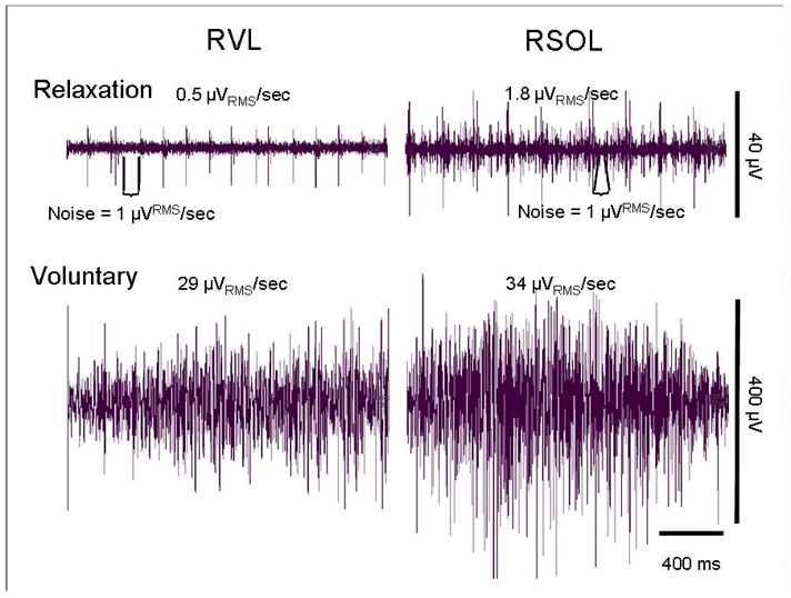
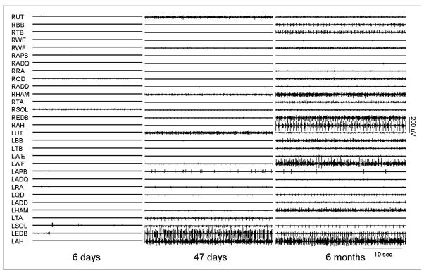
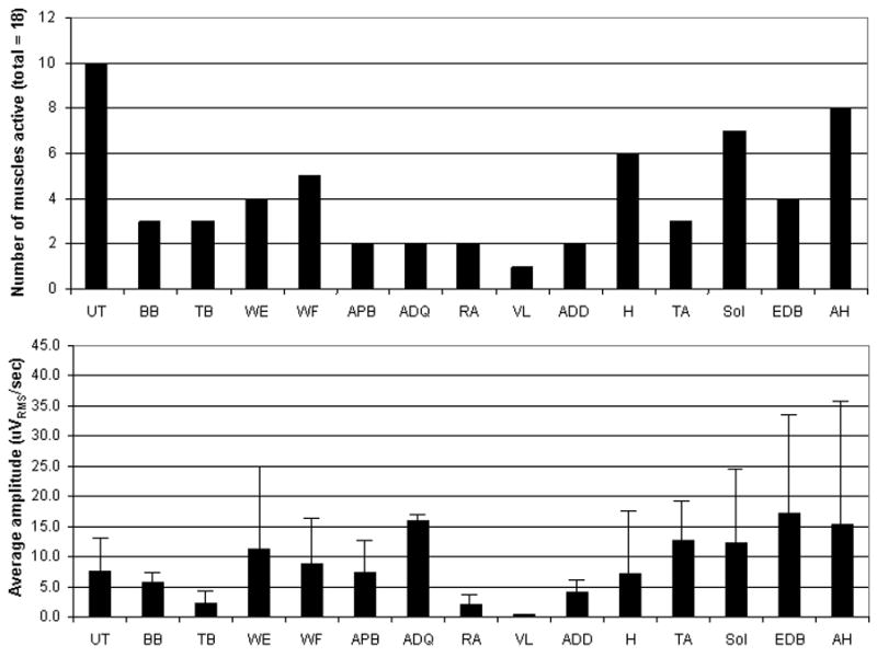
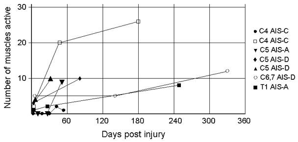
Results: Twelve subjects (48%) showed long-duration involuntary motor-unit activation during relaxation. This activity was seen on initial examination in nine and on follow-up by 3 months post-injury in three others. It was seen in muscles innervated from the injury zone in 11 and caudal to the lesion in 9 subjects. This activity was independent of the presence or absence of tendon reflexes and the ability to volitionally suppress plantar stimulation elicited reflex withdrawal.
Conclusion: The form of involuntary activity described here is the likely result of the altered balance of excitation and inhibition reaching spinal motor neurons because of the loss of inhibitory interneurons or their reduced activation by damaged supraspinal drive and the synaptic reorganization that follows SCI. As such, this activity may be useful for monitoring the effects of neuroprotective and restorative intervention strategies in persons with SCI.
Conflict of interest statement
The authors declare no conflict of interest.
Figures
Similar articles
-
Neurophysiological characterization of motor recovery in acute spinal cord injury.Spinal Cord. 2011 Mar;49(3):421-9. doi: 10.1038/sc.2010.145. Epub 2010 Nov 16. Spinal Cord. 2011. PMID: 21079622 Free PMC article.
-
Biomechanical and electromyographic assessment of spastic hypertonus in motor complete traumatic spinal cord-injured individuals.Spinal Cord. 2011 Jan;49(1):142-8. doi: 10.1038/sc.2010.56. Epub 2010 Jun 8. Spinal Cord. 2011. PMID: 20531358
-
From spinal shock to spasticity: neuronal adaptations to a spinal cord injury.Neurology. 2000 Apr 25;54(8):1574-82. doi: 10.1212/wnl.54.8.1574. Neurology. 2000. PMID: 10762496 Clinical Trial.
-
Human spinal cord injury: motor unit properties and behaviour.Acta Physiol (Oxf). 2014 Jan;210(1):5-19. doi: 10.1111/apha.12153. Epub 2013 Sep 13. Acta Physiol (Oxf). 2014. PMID: 23901835 Review.
-
Mechanisms of Different Motor Neurons in the Occurrence of Spasticity After Spinal Cord Injury: A Narrative Review.Int J Mol Sci. 2025 May 28;26(11):5162. doi: 10.3390/ijms26115162. Int J Mol Sci. 2025. PMID: 40507976 Free PMC article. Review.
Cited by
-
Electromyography-Force Relation and Muscle Fiber Conduction Velocity Affected by Spinal Cord Injury.Bioengineering (Basel). 2023 Feb 6;10(2):217. doi: 10.3390/bioengineering10020217. Bioengineering (Basel). 2023. PMID: 36829711 Free PMC article.
-
Characterizing a Common Class of Spontaneous Movements.Int J Ther Massage Bodywork. 2022 Sep 1;15(3):4-17. doi: 10.3822/ijtmb.v15i3.719. eCollection 2022 Sep. Int J Ther Massage Bodywork. 2022. PMID: 36061228 Free PMC article.
-
Interaction between muscle tone, short-range stiffness and increased sensory feedback gains explains key kinematic features of the pendulum test in spastic cerebral palsy: A simulation study.PLoS One. 2018 Oct 18;13(10):e0205763. doi: 10.1371/journal.pone.0205763. eCollection 2018. PLoS One. 2018. PMID: 30335860 Free PMC article.
-
Transcutaneous spinal cord stimulation and motor responses in individuals with spinal cord injury: A methodological review.PLoS One. 2021 Nov 18;16(11):e0260166. doi: 10.1371/journal.pone.0260166. eCollection 2021. PLoS One. 2021. PMID: 34793572 Free PMC article.
-
Wiener filtering of surface EMG with a priori SNR estimation toward myoelectric control for neurological injury patients.Med Eng Phys. 2014 Dec;36(12):1711-5. doi: 10.1016/j.medengphy.2014.09.008. Epub 2014 Oct 29. Med Eng Phys. 2014. PMID: 25443536 Free PMC article.
References
-
- Mahoney JS, Engebretson JC, Cook KF, Hart KA, Robinson-Whelen S, Sherwood AM. Spasticity experience domains in persons with spinal cord injury. Arch Phys Med Rehabil. 2007;88:287–94. - PubMed
-
- Adams AA, Hicks AL. Spasticity after spinal cord injury. Spinal Cord. 2005;43:577–586. - PubMed
-
- Sherwood AM, McKay WB. Assessment of spasticity and upper motor neuron dysfunction. In: Akay Metin., editor. Wiley Encyclopedia of Biomedical Engineering. Vol. 5. John Wiley & Sons, Inc; 2006. pp. 3306–3315.
-
- Sherwood AM, McKay WB, Dimitrijevic MR. Motor control after spinal cord injury: assessment using surface EMG. Muscle Nerve. 1996;19(8):966–79. - PubMed
Publication types
MeSH terms
Grants and funding
LinkOut - more resources
Full Text Sources
Medical
Research Materials
