Review
. 2010 Aug;6(8):453-60.
doi: 10.1038/nrrheum.2010.102. Epub 2010 Jun 29.

Cellular and molecular aspects of vascular dysfunction in systemic sclerosis

Affiliations
Review

Cellular and molecular aspects of vascular dysfunction in systemic sclerosis

Maria Trojanowska. Nat Rev Rheumatol. 2010 Aug.

Abstract

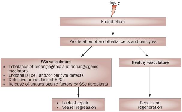
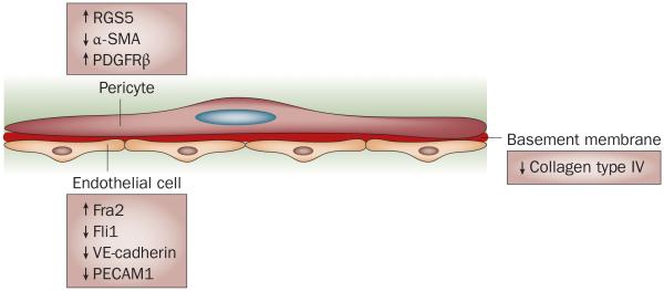
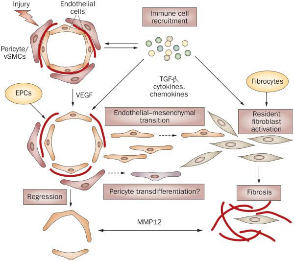
Systemic sclerosis (SSc) is characterized by vascular alterations, activation of the immune system and tissue fibrosis. Vascular insufficiency manifests early in the disease, and although there is evidence of an active repair process, capillaries deteriorate and regress. Factors that contribute to the failure of vascular regeneration might include persistent injury, an imbalance between proangiogenic and antiangiogenic mediators, intrinsic abnormal properties of the cellular components of the vessels, and the presence of fibroblast-derived antiangiogenic factors. In addition, circulating dysfunctional endothelial progenitor cells might further exacerbate vessel deterioration. Abnormal expression of transcription factors, including Fra2 and Fli1, has been proposed to contribute to SSc vasculopathy. Fli1 regulates genes that are involved in vessel maturation and stabilization, suggesting that reduced levels of Fli1 in SSc vasculature could contribute to the development of unstable vessels that are prone to regression. Conversely, proliferating endothelial cells and pericytes, in the presence of an appropriate stimulus, might transdifferentiate into collagen-producing cells, and thus contribute to the initiation of fibrosis. Despite progress in treating the symptoms of vascular disease in SSc, the underlying mechanisms remain poorly understood. An improved knowledge of the molecular and cellular pathways that contribute to SSc vasculopathy could help in the design of effective therapies in the future.


Figures

Figure 1
Figure 1
Schematic overview of the principal stages of angiogenesis. (1) In quiescent capillaries, endothelial cells form tight contacts with each other and with neighboring pericytes. Both cell types are embedded in the shared basement membrane. (2) Angiogenic sprouting requires local degradation of extracellular components of the basement membrane by endothelial-cell-derived proteases, loosening of the cell–cell and cell–matrix contacts, endothelial cell proliferation, migration, and formation of a nonlumenized sprout. Factors secreted by the endothelial cells attract pericytes that contribute to stabilization of the newly formed vessel. (3) Maturation of capillaries involves re-establishment of cell–cell contacts, lumen formation, secretion of new basement membrane, and recruitment of pericytes.
Figure 2
Figure 2
Schematic overview of the potential mechanisms involved in impaired neoangiogenesis in SSc. In healthy individuals, injury to the endothelium triggers a repair process that leads to vessel regeneration. In patients with SSc, there is evidence that endothelial cells and pericytes proliferate during the early stages of the disease, suggesting an initiation of the angiogenic process in response to injury. However, the repair process fails, ultimately leading to regression of small vessels. Studies have revealed several potential mechanisms that could account for the impairment of the angiogenic process in SSc, but the specific contribution of each of these mechanisms to SSc vasculopathy requires further evaluated. Abbreviations: EPCs, endothelial progenitor cells; SSc, systemic sclerosis.
Figure 3
Figure 3
Molecular alterations of components of the SSc vasculature. Pericytes exhibit markers of immaturity or proliferation, including upregulation of RGS5 and PDGFRβ and downregulation of α-SMA. Endothelial cells are characterized by an increased level of the proapoptotic transcription factor Fra2, and decreased levels of the Fli1 transcription factor, VE-cadherin and PECAM1, which are indicative of impaired cell–cell contact, indicating a potential contribution to vessel leakiness. The levels of collagen type IV are reduced, suggesting alterations in the composition of the basement membrane. Abbreviations: α-SMA, α-smooth muscle actin; PDGFRβ, platelet-derived growth factor β receptor; PECAM1, platelet endothelial cell adhesion molecule 1; RGS5, regulator of G protein signaling 5; SSc, systemic sclerosis; VE-cadherin, vascular endothelial cadherin.
Figure 4
Figure 4
Possible links between vasculopathy and fibrosis in SSc. Vascular injury due to an unknown trigger and the influx of the immune cells are the early events in SSc. High levels of VEGF and other proangiogenic mediators derived from the activated immune cells facilitate proliferation of endothelial cells and pericytes in an attempt to restore injured vessels. For reasons that are still not clearly understood, but which may involve imbalance of proangiogenic and antiangiogenic mediators, intrinsic properties of endothelial cells, and the fibroblast-secreted antiangiogenic factors (for example, MMP12) this process fails, leading to vessel regression. In the presence of TGF-β or other immune mediators (such as cytokines and chemokines), or both, endothelial cells could acquire a migratory phenotype through endothelial–mesenchymal transition and enter the surrounding tissue, where they further differentiate into collagen-producing cells. Likewise, pericytes could transdifferentiate into fibroblasts or myofibroblasts and produce collagen. In SSc, intrinsic abnormal properties of endothelial cells and pericytes might render vascular cells particularly susceptible to undergoing these transitions. Activated resident fibroblasts and fibrocytes that enter the injured tissue from the circulation are likely to represent additional source of collagen producing cells that contribute to fibrosis in SSc lesion. Abbreviations: EPCs, endothelial progenitor cells; VEGF, vascular endothelial growth factor; MMP12, matrix metalloproteinase 12; SSc, systemic sclerosis; TGF-β, transforming growth factor β.

References

    1. Sapadin AN, Esser AC, Fleischmajer R. Immunopathogenesis of scleroderma—evolving concepts. Mt Sinai J. Med. 2001;68:233–242. - PubMed
    1. Varga J, Abraham D. Systemic sclerosis: a prototypic multisystem fibrotic disorder. J. Clin. Invest. 2007;117:557–567. - PMC - PubMed
    1. Bakst R, Merola JF, Franks AG, Jr, Sanchez M. Raynaud’s phenomenon: pathogenesis and management. J. Am. Acad. Dermatol. 2008;59:633–653. - PubMed
    1. LeRoy EC, Medsger TA., Jr. Raynaud’s phenomenon: a proposal for classification. Clin. Exp. Rheumatol. 1992;10:485–488. - PubMed
    1. Koenig M, et al. Autoantibodies and microvascular damage are independent predictive factors for the progression of Raynaud’s phenomenon to systemic sclerosis: a twenty-year prospective study of 586 patients, with validation of proposed criteria for early systemic sclerosis. Arthritis Rheum. 2008;58:3902–3912. - PubMed

MeSH terms