

doi: 10.1007/s12551-008-0006-z.
Epub 2009 Jan 17.
Phase-plate electron microscopy: a novel imaging tool to reveal close-to-life nano-structures
Affiliations
- PMID: 20585379
- PMCID: PMC2883085
- DOI: 10.1007/s12551-008-0006-z
Item in Clipboard
Phase-plate electron microscopy: a novel imaging tool to reveal close-to-life nano-structures
Biophys Rev.
2009 Mar.
Abstract
After slow progress in the efforts to develop phase plates for electron microscopes, functional phase plate using thin carbon film has been reported recently. It permits collecting high-contrast images of close-to-life biological structures with cryo-fixation and without staining. This report reviews the state of the art for phase plates and what is innovated with them in biological electron microscopy. The extension of thin-film phase plates to the material-less type using electrostatic field or magnetic field is also addressed. ELECTRONIC SUPPLEMENTARY MATERIAL: The online version of this article (doi:10.1007/s12551-008-0006-z) contains supplementary material, which is available to authorized users.
Figures
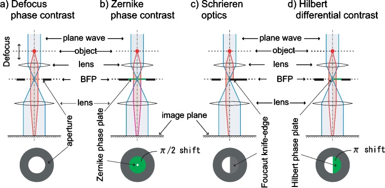
Various phase plates a A spatial filter schematic for the defocus phase contrast (DPC). b A spatial filter schematic for the Zernike phase contrast (ZPC). c A spatial filter schematic for Schlieren optics (SO) or single-sideband imaging (SSI). d A spatial filter schematic for Hilbert differential contrast (HDC)

TEM images (300 kV) for proteins and viruses. a A ZPC image for an ice-embedded sample of GroEL (Danev and Nagayama 2008). The inset is a diffractogram obtained by Fourier-transform of the image. A typical cosine CTF is shown. This can be experimentally determined with the kind of diffractogram shown in the inset. b A conventional image for an ice-embedded sample of GroEL (Danev and Nagayama 2008). The inset is a diffractogram obtained by Fourier-transform of the image. A typical sine CTF is shown. This can be experimentally determined with the kind of diffractogram shown in the inset. c A ZPC image for a tubulate membrane liposome wrapped by a polymeric protein fiber (Shimada et al. 2007). d A ZPC image of ice-embedded influenza A viruses (Yamaguchi et al. 2008)
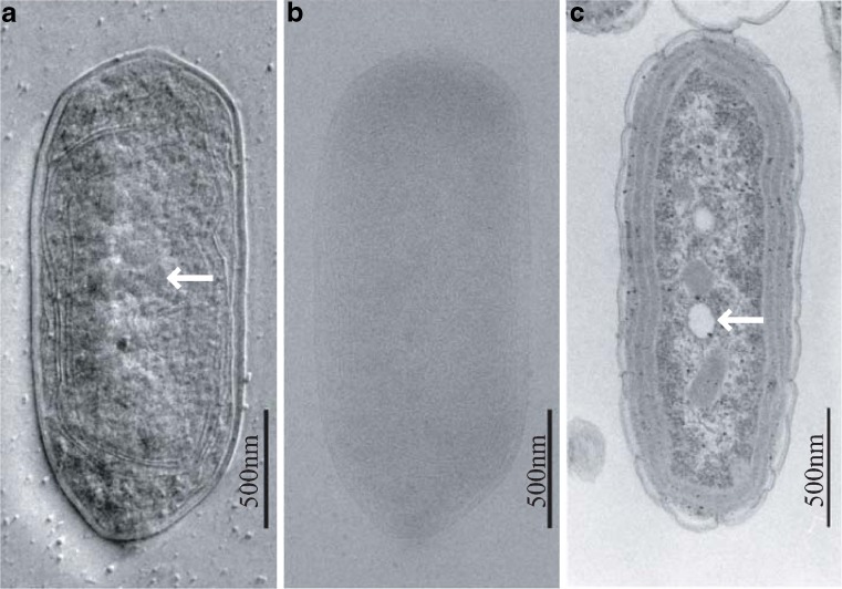
TEM images (300 kV) of a vitrified cyanobacterial cell. a A HDC just-focused image of an ice-embedded cyanobacterial whole cell (Kaneko et al. 2005). The polyphosphate bodies (shown by an arrow) are prominent among detailed ultrastructures. b A conventional TEM image of an ice-embedded cyanobacterial cell for the same view field as a defocus setting of 15 μm (Kaneko et al. 2005). c A 100 kV conventional TEM image of a chemically fixed, resin-embedded, and thin-sectioned cell stained with uranyl acetate and lead citrate (Kaneko et al. 2005). Polyphosphate bodies are lost during the preparation process. This leaves an empty hole in the section (arrow) (Kaneko et al. 2005)

A three-layered carbon film design to avoid the charging effect in a Zernike phase plate (Nagayama 2005). Contaminants are wrapped by a conductive carbon coat to shield the electrostatic potential

a An electrostatic phase plate made of a five-layered electrode supported by three spokes (Majorovits et al. 2007). b An electrostatic phase plate made of a double-layered electrode supported by a single spoke (Cambie et al. 2007). c An example of a five-bar magnet (0.8 μm width and 2 mm length) made of cobalt thin-film deposited on a biprism (Nagayama 2008). d A schematic of an anamorphotic phase plate. The spit-shaped device has embedded electrodes for application of electrostatic fields to a high aspect ratio anamorphotic diffraction plane electron wave (Schröder et al. 2007)
References
-
- Aharonov Y, Bohm D. Significance of electromagnetic potentials in the quantum theory. Phys Rev. 1959;115:485–491. doi: 10.1103/PhysRev.115.485. - DOI
-
- Boersch H. Über die Kontraste von Atomen in Electronenmikroskop. Z Naturforschg. 1947;2a:615–633.
LinkOut - more resources
Full Text Sources
Other Literature Sources
