. 2010 Jun 18;5(6):e11199.
doi: 10.1371/journal.pone.0011199.

Immune response to Lactobacillus plantarum expressing Borrelia burgdorferi OspA is modulated by the lipid modification of the antigen

Affiliations

Immune response to Lactobacillus plantarum expressing Borrelia burgdorferi OspA is modulated by the lipid modification of the antigen

Beatriz del Rio et al. PLoS One. .

Abstract

Background: Over the past decade there has been increasing interest in the use of lactic acid bacteria as mucosal delivery vehicles for vaccine antigens, microbicides and therapeutics. We investigated the mechanism by which a mucosal vaccine based in recombinant lactic acid bacteria breaks the immunological tolerance of the gut in order to elicit a protective immune response.

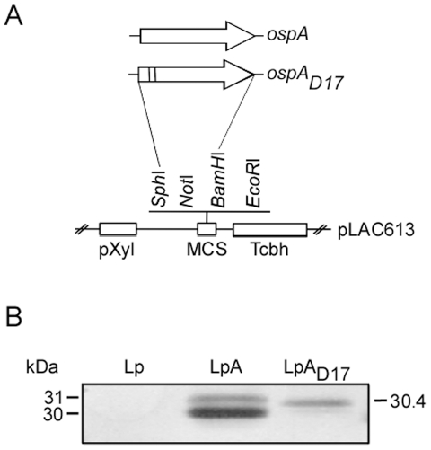
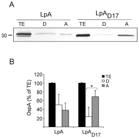
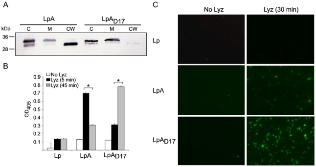
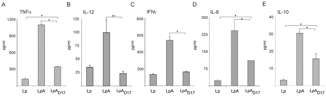
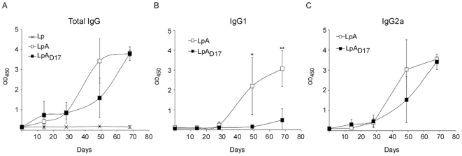
Methodology/principal findings: We analyzed how the lipid modification of OspA affects the localization of the antigen in our delivery vehicle using a number of biochemistry techniques. Furthermore, we examined how OspA-expressing L. plantarum breaks the oral tolerance of the gut by stimulating human intestinal epithelial cells, peripheral blood mononuclear cells and monocyte derived dendritic cells and measuring cytokine production. We show that the leader peptide of OspA targets the protein to the cell envelope of L. plantarum, and it is responsible for protein export across the membrane. Mutation of the lipidation site in OspA redirects protein localization within the cell envelope. Further, we show that lipidated-OspA-expressing L. plantarum does not induce secretion of the pro-inflammatory cytokine IL-8 by intestinal epithelial cells. In addition, it breaks oral tolerance of the gut via Th1/Th2 cell mediated immunity, as shown by the production of pro- and anti-inflammatory cytokines by human dendritic cells, and by the production of IgG2a and IgG1 antibodies, respectively.

Conclusions/significance: Lipid modification of OspA expressed in L. plantarum modulates the immune response to this antigen through a Th1/Th2 immune response.


Conflict of interest statement

Competing Interests: The corresponding author (MGS) has a relevant patent and is a major stockholder in Biopeptides, Corp.; JFMLS has a relevant patent and is a major stockholder in Lactrys Biopharmaceuticals BV; Patent title: Live Bacterial Vaccine. Patent number US12/304,634 filling date 03/31/2009. The authors confirm that this does not alter their adherence to all the PLoS ONE policies on sharing data and materials, as detailed online in PLoS guide for authors. BD does not have any potential financial conflict of interest related to this manuscript.

Figures

Figure 1
Figure 1. Characterization of recombinant Lactobacillus.
Schematic representation of the ospA and ospAD17 constructs (A), and immunoblot characterization (B). Whole-cell extract of control, OspA- and OspAD17-expressing L. plantarum (Lp, LpA, and LpAD17, respectively) were analyzed on a 12% SDS-PAGE, transferred to PVDF membrane and tested with OspA-specific monoclonal antibody 184.1. Legend: pXyl, xylose promoter; MCS, multicloning site; Tcbh, Rho independent transcriptional terminator.
Figure 2
Figure 2. Evaluation of protein export and lipidation.
OspA- and OspAD17-expressing L. plantarum were disrupted with a French® press, the insoluble material (cell envelope) was extracted with Triton X-114 and partitioned into detergent and aqueous phases. Protein fractions were analyzed on a SDS-PAGE and tested by immunoblot with OspA-specific monoclonal antibody LA2.2 (A). Protein was quantified by densitometry. The results were plotted as a percentage of the total OspA content for each recombinant Lactobacillus (B). TE, total extract; D, detergent phase; A, aqueous phase; OspA- and OspAD17-expressing L. plantarum (LpA and LpAD17, respectively). *p<0.05.
Figure 3
Figure 3. Localization of recombinant antigens in L. plantarum.
Localization of the recombinant antigens was studied by (A) serial cell fractionation, (B) live-cell ELISA (lcELISA) and (C) Immunofluorescence Assay (IFA). (A) Immunoblot of cellular fractions of L. plantarum expressing OspA antigens: C, cytosol; M, membrane; CW, cell wall. L. plantarum was treated with 250 kU/ml Lysozyme (Lyz) for 45 min to digest the cell wall; protoplasts were disrupted and, membrane and cell wall fractions were separated by ultracentrifugation. 3 µg of each fraction was analyzed in a 12% SDS-PAGE, transferred to PVDF membrane, and tested by immunoblot with OspA-specific monoclonal antibody LA2.2 (mAb LA2.2). (B) Live recombinant L. plantarum were treated during 0, 5 or 45 min with Lyz and then subjected to lcELISA using mAb LA2.2 and anti-mouse IgG secondary antibody labeled with alkaline phosphatase. The Optical Density at 405 nm (OD405) of the mean endpoint titer was determined. The average of triplicate samples per sample was determined and the error bar indicates standard deviation. (C) Live recombinant L. plantarum were treated with or without Lyz for 30 min. After cell wall removal, the cells were incubated with mAb LA2.2 followed by Alexa Fluor 488-labeled goat anti-mouse IgG (1∶250) antibodies. Immunofluorescence staining was visualized using a Zeiss inverted Axiovert 200 microscope, and the images were acquired using AxioVision software. *p<0.001. Results are representative of one of three independent experiments.
Figure 4
Figure 4. Production of IL-8 in human epithelial cells co-cultured with recombinant L. plantarum.
Human epithelial cells (T84) were seeded in 24-well plates (106 cells/well) and grown until they reached about 95% confluence. UV-killed L. plantarum expressing OspA (LpA) or the mutant OspAD17 (LpAD17) were co-cultured with T84 cells at MOI 10:1 colony-forming units per cell for 48 h and culture supernatants were collected to determine IL-8 secretion by sandwich ELISA (Quantikine). 0.5 µg/ml TNFα and UV-killed L. plantarum (Lp) were used as positive and negative control, respectively. The average of triplicate samples was determined and the error bar indicates standard deviation. Results are representative of one of three independent experiments.
Figure 5
Figure 5. Production of cytokines in human MDDCs co-cultured with purified recombinant wtOspA or mutOspA.
Monocytes were isolated from human PBMCs by MACS using the Monocyte Isolation Kit II. Monocytes were derived to immature DCs (MDDCs) by cultivating the cells in the presence of 100 nM GM-CSF and 10 nM IL-4 for 5 days. MDDCs (2×10⁵ cells/well) were seed in 24 well plates and co-cultured with 2.5 µg/ml of wtOspA or mutOspA. After 48 h of stimulation, supernatants were collected and TNFα (A), IL-12 (B), IFNγ (C) and IL-10 (D) cytokine production was measured by sandwich ELISA (Quantikine). *p<0.001,**p<0.05. Results are representative of one of three independent experiments.
Figure 6
Figure 6. Production of cytokines in human PBMC/DCs co-cultured with recombinant Lactobacillus.
Human Peripheral Blood Mononuclear Cells (PBMCs) were treated with 100 nM GM-CSF and 10 nM IL-4 during 5 days to derive monocytes into dendritic cells. 106 cells/well were seed in 24 well plates and co-cultured with UV-killed recombinant Lactobacillus expressing OspA (LpA) or the mutant OspAD17 (LpAD17) at MOI 10:1 colony-forming units per cell. 100 ng/ml Escherichia coli O111:B4 lipopolysaccharide (LPS) and L. plantarum (Lp) were used as positive and negative control, respectively. After 48 h of stimulation, supernatants were collected and TNFα (A), IL-12 (B), IFNγ (C), IL-6 (D) and IL-10 (E) cytokine production was measured by sandwich ELISA (Quantikine). *p<0.001,**p<0.02. Results are representative of one of three independent experiments.
Figure 7
Figure 7. Production of cytokines in human MDDC co-cultured with recombinant Lactobacillus.
Monocytes were isolated from human PBMCs by MACS using the Monocyte Isolation Kit II. Monocytes were derived to immature DCs (MDDCs) by cultivating the cells in the presence of 100 nM GM-CSF and 10 nM IL-4 for 5 days. MDDCs (2×10⁵ cells/well) were co-cultured with UV-killed recombinant Lactobacillus expressing OspA (LpA) or the mutant OspAD17 (LpAD17) at MOI 10:1 colony-forming units per DC. 100 ng/ml Escherichia coli O111:B4 lipopolysaccharide (LPS) and L. plantarum (Lp) were used as positive and negative control, respectively. After 48 h of stimulation, supernatants were collected and TNF (A), IL-12 (B), IFNγ (C), IL-6 (D) and IL-10 (E) cytokine production was measured by sandwich ELISA (Quantikine). *p<0.05,**p<0.001. Results are representative of one of three independent experiments.
Figure 8
Figure 8. Antibody response to oral administration of recombinant L. plantarum: serological IgG.
C3H-HeJ mice were inoculated intragastrically with L. plantarum expressing OspA (LpA) or the mutant OspAD17 (LpAD17). Control mice were inoculated with L. plantarum (Lp). Serum samples were collected at days 0, 14, 28, 49 and 68, and specific serological anti-OspA total IgG (A), IgG1 (B) and IgG2a (C) antibodies were measured by indirect ELISA. The results are expressed as Optical Density at 450 nm (OD450). The average of triplicate samples per mouse was determined and the error bar indicates standard deviation. *p<0.05, **p<0.01. Results are representative of one of three independent experiments. n = 4 mice per group.
Figure 9
Figure 9. Antibody response to oral administration of recombinant L. plantarum: mucosal IgA.
C3H-HeJ mice were inoculated intragastrically with L. plantarum expressing OspA (LpA) or the mutant OspAD17 (LpAD17). Control mice were inoculated with L. plantarum (Lp). Specific mucosal anti-OspA IgA antibodies were measured by indirect ELISA in broncheoalveolar lavage (BAL) (A) and stool (B) collected on day 68. The results corresponding to each mouse are expressed as Optical Density at 450 nm (OD450) of the mean endpoint titer. Results are representative of one of three independent experiments. n = 4 mice per group. ns, not significant.

References

    1. Wells JM, Mercenier A. Mucosal delivery of therapeutic and prophylactic molecules using lactic acid bacteria. Nat Rev Microbiol. 2008;6:349–362. - PMC - PubMed
    1. Anderson R, Dougan G, Roberts M. Delivery of the Pertactin/P.69 polypeptide of Bordetella pertussis using an attenuated Salmonella typhimurium vaccine strain: expression levels and immune response. Vaccine. 1996;14:1384–1390. - PubMed
    1. Ascon MA, Hone DM, Walters N, Pascual DW. Oral immunization with a Salmonella typhimurium vaccine vector expressing recombinant enterotoxigenic Escherichia coli K99 fimbriae elicits elevated antibody titers for protective immunity. Infect Immun. 1998;66:5470–5476. - PMC - PubMed
    1. Kohler JJ, Pathangey L, Hasona A, Progulske-Fox A, Brown TA. Long-term immunological memory induced by recombinant oral Salmonella vaccine vectors. Infect Immun. 2000;68:4370–4373. - PMC - PubMed
    1. Peters C, Peng X, Douven D, Pan ZK, Paterson Y. The induction of HIV Gag-specific CD8+ T cells in the spleen and gut-associated lymphoid tissue by parenteral or mucosal immunization with recombinant Listeria monocytogenes HIV Gag. J Immunol. 2003;170:5176–5187. - PubMed

Publication types

MeSH terms