

Role of Abl kinase and the Wave2 signaling complex in HIV-1 entry at a post-hemifusion step
- PMID: 20585556
- PMCID: PMC2887473
- DOI: 10.1371/journal.ppat.1000956
Role of Abl kinase and the Wave2 signaling complex in HIV-1 entry at a post-hemifusion step
Abstract
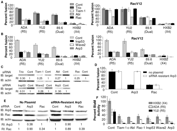
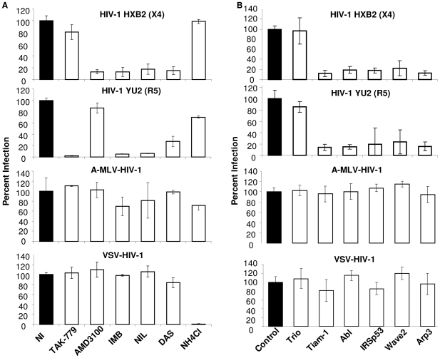
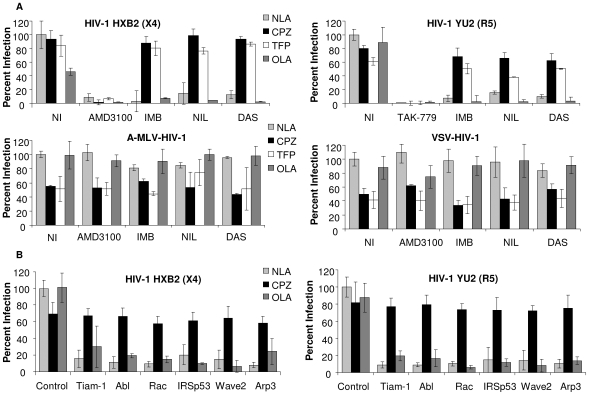
Entry of human immunodeficiency virus type 1 (HIV-1) commences with binding of the envelope glycoprotein (Env) to the receptor CD4, and one of two coreceptors, CXCR4 or CCR5. Env-mediated signaling through coreceptor results in Galphaq-mediated Rac activation and actin cytoskeleton rearrangements necessary for fusion. Guanine nucleotide exchange factors (GEFs) activate Rac and regulate its downstream protein effectors. In this study we show that Env-induced Rac activation is mediated by the Rac GEF Tiam-1, which associates with the adaptor protein IRSp53 to link Rac to the Wave2 complex. Rac and the tyrosine kinase Abl then activate the Wave2 complex and promote Arp2/3-dependent actin polymerization. Env-mediated cell-cell fusion, virus-cell fusion and HIV-1 infection are dependent on Tiam-1, Abl, IRSp53, Wave2, and Arp3 as shown by attenuation of fusion and infection in cells expressing siRNA targeted to these signaling components. HIV-1 Env-dependent cell-cell fusion, virus-cell fusion and infection were also inhibited by Abl kinase inhibitors, imatinib, nilotinib, and dasatinib. Treatment of cells with Abl kinase inhibitors did not affect cell viability or surface expression of CD4 and CCR5. Similar results with inhibitors and siRNAs were obtained when Env-dependent cell-cell fusion, virus-cell fusion or infection was measured, and when cell lines or primary cells were the target. Using membrane curving agents and fluorescence microscopy, we showed that inhibition of Abl kinase activity arrests fusion at the hemifusion (lipid mixing) step, suggesting a role for Abl-mediated actin remodeling in pore formation and expansion. These results suggest a potential utility of Abl kinase inhibitors to treat HIV-1 infected patients.
Conflict of interest statement
The authors have declared that no competing interests exist.
Figures


Similar articles
-
HIV-1 triggers WAVE2 phosphorylation in primary CD4 T cells and macrophages, mediating Arp2/3-dependent nuclear migration.J Biol Chem. 2014 Mar 7;289(10):6949-6959. doi: 10.1074/jbc.M113.492132. Epub 2014 Jan 10. J Biol Chem. 2014. PMID: 24415754 Free PMC article.
-
Induction of the Galpha(q) signaling cascade by the human immunodeficiency virus envelope is required for virus entry.J Virol. 2008 Sep;82(18):9191-205. doi: 10.1128/JVI.00424-08. Epub 2008 Jul 16. J Virol. 2008. PMID: 18632858 Free PMC article.
-
Fusion Stage of HIV-1 Entry Depends on Virus-Induced Cell Surface Exposure of Phosphatidylserine.Cell Host Microbe. 2017 Jul 12;22(1):99-110.e7. doi: 10.1016/j.chom.2017.06.012. Cell Host Microbe. 2017. PMID: 28704658 Free PMC article.
-
HIV coreceptors: from discovery and designation to new paradigms and promise.Eur J Med Res. 2007 Oct 15;12(9):375-84. Eur J Med Res. 2007. PMID: 17933717 Review.
-
Molecular Mechanism of HIV-1 Entry.Trends Microbiol. 2019 Oct;27(10):878-891. doi: 10.1016/j.tim.2019.06.002. Epub 2019 Jun 28. Trends Microbiol. 2019. PMID: 31262533 Free PMC article. Review.
Cited by
-
The membrane fusion step of vaccinia virus entry is cooperatively mediated by multiple viral proteins and host cell components.PLoS Pathog. 2011 Dec;7(12):e1002446. doi: 10.1371/journal.ppat.1002446. Epub 2011 Dec 15. PLoS Pathog. 2011. PMID: 22194690 Free PMC article.
-
HIV-1 triggers WAVE2 phosphorylation in primary CD4 T cells and macrophages, mediating Arp2/3-dependent nuclear migration.J Biol Chem. 2014 Mar 7;289(10):6949-6959. doi: 10.1074/jbc.M113.492132. Epub 2014 Jan 10. J Biol Chem. 2014. PMID: 24415754 Free PMC article.
-
Molecular determinants of the ratio of inert to infectious virus particles.Prog Mol Biol Transl Sci. 2015;129:285-326. doi: 10.1016/bs.pmbts.2014.10.012. Epub 2014 Dec 1. Prog Mol Biol Transl Sci. 2015. PMID: 25595808 Free PMC article. Review.
-
Actin-regulated Siglec-1 nanoclustering influences HIV-1 capture and virus-containing compartment formation in dendritic cells.Elife. 2023 Mar 20;12:e78836. doi: 10.7554/eLife.78836. Elife. 2023. PMID: 36940134 Free PMC article.
-
Role of Drebrin at the Immunological Synapse.Adv Exp Med Biol. 2017;1006:271-280. doi: 10.1007/978-4-431-56550-5_15. Adv Exp Med Biol. 2017. PMID: 28865025 Free PMC article. Review.
References
Publication types
MeSH terms
Substances
Grants and funding
LinkOut - more resources
Full Text Sources
Other Literature Sources
Medical
Research Materials
Miscellaneous
