. 2010 Jun 17;6(6):e1000968.
doi: 10.1371/journal.ppat.1000968.

Complement receptor 1 is a sialic acid-independent erythrocyte receptor of Plasmodium falciparum

Affiliations

Complement receptor 1 is a sialic acid-independent erythrocyte receptor of Plasmodium falciparum

Carmenza Spadafora et al. PLoS Pathog. .

Abstract

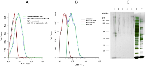
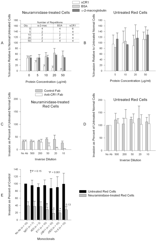
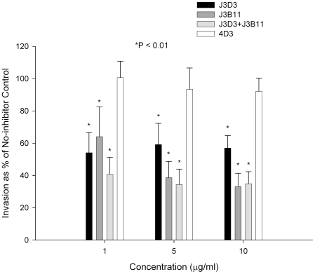
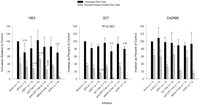
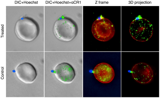
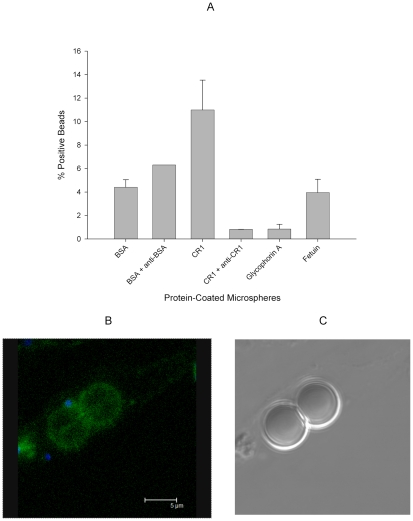
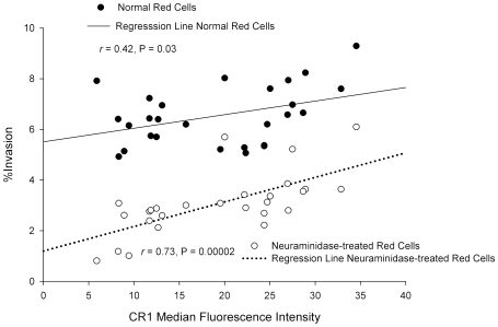
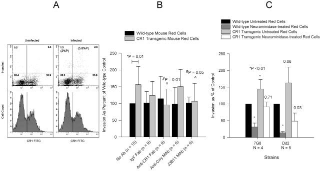
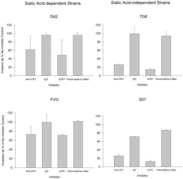
Plasmodium falciparum is a highly lethal malaria parasite of humans. A major portion of its life cycle is dedicated to invading and multiplying inside erythrocytes. The molecular mechanisms of erythrocyte invasion are incompletely understood. P. falciparum depends heavily on sialic acid present on glycophorins to invade erythrocytes. However, a significant proportion of laboratory and field isolates are also able to invade erythrocytes in a sialic acid-independent manner. The identity of the erythrocyte sialic acid-independent receptor has been a mystery for decades. We report here that the complement receptor 1 (CR1) is a sialic acid-independent receptor for the invasion of erythrocytes by P. falciparum. We show that soluble CR1 (sCR1) as well as polyclonal and monoclonal antibodies against CR1 inhibit sialic acid-independent invasion in a variety of laboratory strains and wild isolates, and that merozoites interact directly with CR1 on the erythrocyte surface and with sCR1-coated microspheres. Also, the invasion of neuraminidase-treated erythrocytes correlates with the level of CR1 expression. Finally, both sialic acid-independent and dependent strains invade CR1 transgenic mouse erythrocytes preferentially over wild-type erythrocytes but invasion by the latter is more sensitive to neuraminidase. These results suggest that both sialic acid-dependent and independent strains interact with CR1 in the normal red cell during the invasion process. However, only sialic acid-independent strains can do so without the presence of glycophorin sialic acid. Our results close a longstanding and important gap in the understanding of the mechanism of erythrocyte invasion by P. falciparum that will eventually make possible the development of an effective blood stage vaccine.


Conflict of interest statement

The authors have declared that no competing interests exist.

Figures

Figure 1
Figure 1. Chicken anti-human CR1 recognizes red cell CR1.
(A) Binding of chicken anti-human CR1 Fab is abolished by treatment of erythrocytes with trypsin but not with neuraminidase. (B) Binding of chicken anti-CR1 Fab to human red cells is abolished by incubation with soluble CR1 (sCR1) but not by bovine serum albumin. (C) PAGE of chicken anti-CR1 immunoprecipitates, sCR1 and red cell lysates run on Novex Nupage Bis-tris 4–12% acrylamide gel under reducing conditions and stained with silver. Lane 1: Immunoprecipitate with chicken anti-CR1 from intact red cell lysate. Lane 2: Immunoprecipitate with chicken anti-CR1 from trypsin-treated red cell lysate. Lane 3: Immunoprecipitate with control chicken IgY from intact red cell lysate. Lane 4: Immunoprecipitate with chicken IgY from trypsin-treated red cell lysate. Lane 5: 0.01 µg of purified sCR1. Lane 6: 1 µl of intact red cell lysate used for immunoprecipitation. Lane 7: 1 µl of trypsin-treated red cell lysate used for immunoprecipitation.
Figure 2
Figure 2. Inhibition of P. falciparum 7G8 invasion by sCR1 and antibodies against CR1.
(A) sCR1 blocked invasion of neuraminidase-treated erythrocytes in a dose-dependent manner while bovine serum albumin (BSA) and α-2-macroglobulin had no effect. (B) No protein had an effect on the invasion of untreated erythrocytes. (C) Chicken anti-human CR1 Fab blocked invasion of neuraminidase-treated erythrocytes in a dose-dependent manner while purified chicken IgY Fab control had no effect. (D) No antibody had effect on the inhibition of intact red cells. (C–D) The concentration of antibody stocks was 80 µg/ml. Summary of three experiments. (E) Inhibition of P. falciparum 7G8 invasion using anti-CR1 monoclonal antibodies. Numbers above bars are P values for the comparison with the no antibody (No Ab) control using Dunnett's pairwise multiple comparison t-test, taking into account matching by experiment date. An IgG1 irrelevant monoclonal (R&D Systems, Minneapolis, MN, USA) and anti-CD55 monoclonal (clone NaM16-4D3) (4D3) (Santa Cruz Biotechnology, Inc.) were used as negative controls. E11, To5, J3D3, and J3B11 are anti-CR1 monoclonal antibodies. All monoclonal antibodies were used at 1 µg/ml except in two experiments where every monoclonal was used at 15 µg/ml. Bars represent means ± standard deviations. Invasion rates in untreated controls (no antibody) ranged from 2% to 14%.
Figure 3
Figure 3. Effect of combined monoclonals on sialic acid-independent invasion of 7G8.
Monoclonals J3B11 and J3D3 were used singly or in combination. When used in combination, each monoclonal was used at half of the total concentration. 4D3 is a negative control antibody that recognizes red cell CD55. P values are for the comparison to the no-inhibitor control using Dunnett's pairwise multiple comparison t-test using matching by experiment date. Parasitemia was determined using flow cytometry. The figure represents a summary of 3 experiments. Bars are means ± standard deviations.
Figure 4
Figure 4. Inhibition of invasion of P. falciparum strains HB3, 3D7, and Dd2NM.
HB3 invasion of neuraminidase-treated cells was least affected by monoclonal and polyclonal antibodies against CR1. Monoclonal antibodies were used at 4 µg/ml. Fab fragments were used at 8 µg/ml and purified proteins were used at 50 µg/ml. Numbers above bars are P values for the comparison with the no-inhibitor control using Dunnett's pairwise multiple comparison t test, taking into account matching by experiment date. Bars represent means ± standard deviations. Alpha-2-mac = α-2-macroglobulin. Invasion rates for untreated erythrocytes with no inhibitor ranged from 3% to 37%.
Figure 5
Figure 5. Representative examples of interaction between merozoites and CR1 on neuraminidase-treated and untreated control erythrocytes.
CR1 shows a characteristic speckled pattern with aggregation around the merozoite. All negative control antibodies showed no staining. CR1 and glycophorin A were stained with specific antibodies followed by fluorochrome-conjugated secondary antibodies against glycophorin (red) and CR1 (green). Merozoites (blue) were stained with the nucleic acid specific stain (Hoechst 33342). DIC = differential interference contrast.
Figure 6
Figure 6. 7G8 merozoites bind preferentially to sCR1-coated microspheres.
(A) Protein-coated microspheres were incubated overnight with purified late trophozoites and schizonts. Binding was detected by % of spheres positive for Hoechst staining using flow cytometry. Bars represent means ± standard deviations. The figure represents a summary of 2–3 experiments except for the rabbit anti-BSA which was included in one experiment. (B) Fluorescence image of sCR1-coated microspheres showing attached merozoites (blue) and surface sCR1 (green). (C) Differential interference contrast image of microspheres in B.
Figure 7
Figure 7. Correlation between CR1 median fluorescence intensity (MFI) and invasion of erythrocytes by P. falciparum 7G8.
Erythrocytes from 27 healthy donors were used. To control for day-to-day variation the percent invasion as well as the CR1 MFI were normalized to an erythrocyte standard sample that was used in every experiment. The CR1 MFI was re-measured once in 4 samples with extreme values. In 19 donors the invasion assay was repeated 1–3 times. When more than one measurement was done, the mean was used for normalization.
Figure 8
Figure 8. Invasion of wild-type and human CR1 transgenic mouse erythrocytes by P. falciparum.
(A) 7G8 invaded CR1 transgenic mouse erythrocytes preferentially in a mixed culture of wild-type and CR1 transgenic mouse erythrocytes incubated overnight with or without schizonts and analysed by flow cytometry. Upper panels show dot plots of Hoechst negative (uninfected) and positive (infected) erythrocytes. Numbers in parenthesis represent the percent parasitemia (%P). Numbers without parenthesis in each quadrant represent the percent cells from the total population. The lower panels show CR1 fluorescence histograms and the demarcation between CR1 positive (rectangle) and negative erythrocytes. (B) Effect of polyclonal and monoclonal antibodies on the invasion of wild-type and CR1 transgenic mouse erythrocytes by 7G8. Only significant P values are shown. *P is for the comparison between wild-type and CR1 transgenic red cells with no antibody. #P is for the comparison between antibody and no antibody. Invasion in wild-type erythrocytes ranged from 0.2% to 4.5%. (C) Comparison of invasion of wild-type and CR1 transgenic mouse erythrocytes between 7G8 and Dd2. Numbers above bars represent P values for the comparison to the wild-type untreated control. Invasion was measured by flow cytometry and ranged from 0.45% to 5.5% in wild-type untreated erythrocytes. All bars represent means ± standard deviations.
Figure 9
Figure 9. Inhibition of invasion of neuraminidase-treated red cells by sialic acid-dependent and independent strains.
Bars represent means ± standard deviations of the invasion as % of the no-inhibitor control. The figure represents a summary of 2–4 experiments. Parasitemias were determined by flow cytometry.

References

    1. Miller LH, Haynes JD, McAuliffe FM, Shiroishi T, Durocher JR, et al. Evidence for differences in erythrocyte surface receptors for the malarial parasites, Plasmodium falciparum and Plasmodium knowlesi. J Exp Med. 1977;146:277–281. - PMC - PubMed
    1. Deas JE, Lee LT. Competitive inhibition by soluble erythrocyte glycoproteins of penetration by Plasmodium falciparum. Am J Trop Med Hyg. 1981;30:1164–1167. - PubMed
    1. Perkins M. Inhibitory effects of erythrocyte membrane proteins on the in vitro invasion of the human malarial parasite (Plasmodium falciparum) into its host cell. J Cell Biol. 1981;90:563–567. - PMC - PubMed
    1. Pasvol G, Jungery M, Weatherall DJ, Parsons SF, Anstee DJ, et al. Glycophorin as a possible receptor for Plasmodium falciparum. Lancet. 1982;2:947–950. - PubMed
    1. Maier AG, Duraisingh MT, Reeder JC, Patel SS, Kazura JW, et al. Plasmodium falciparum erythrocyte invasion through glycophorin C and selection for Gerbich negativity in human populations. Nat Med. 2003;9:87–92. - PMC - PubMed

Publication types

MeSH terms