. 2010 Jun 17;6(6):e1000954.
doi: 10.1371/journal.ppat.1000954.

A kinome RNAi screen identified AMPK as promoting poxvirus entry through the control of actin dynamics

Affiliations

A kinome RNAi screen identified AMPK as promoting poxvirus entry through the control of actin dynamics

Theresa S Moser et al. PLoS Pathog. .

Abstract

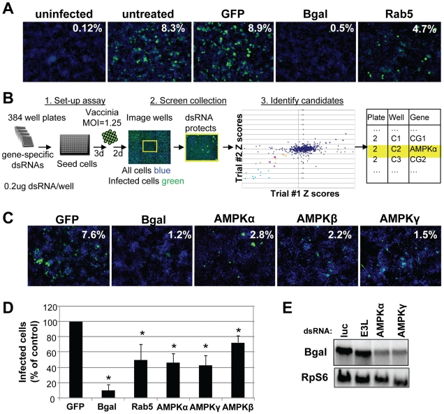
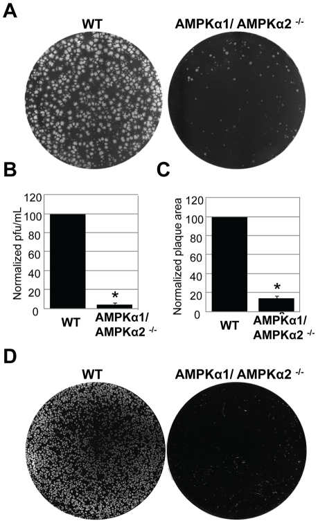
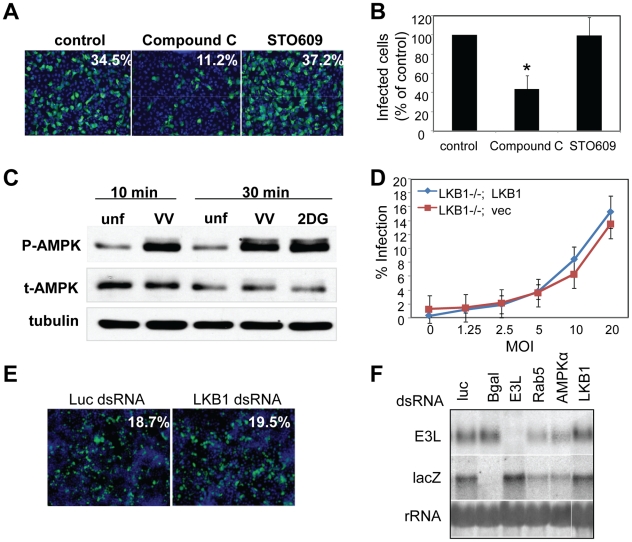
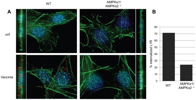
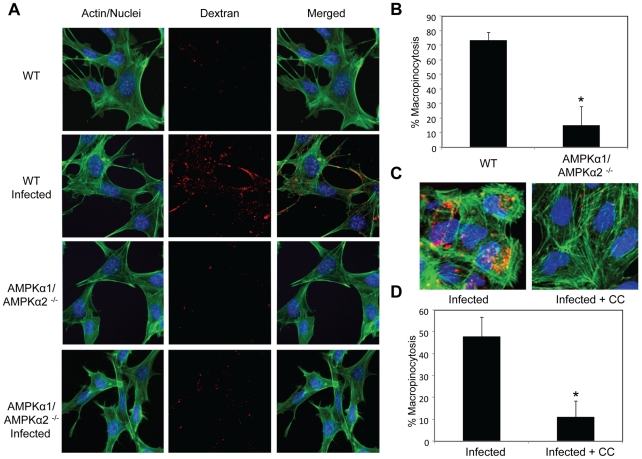
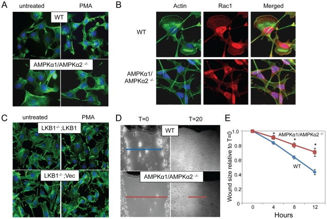
Poxviruses include medically important human pathogens, yet little is known about the specific cellular factors essential for their replication. To identify genes essential for poxvirus infection, we used high-throughput RNA interference to screen the Drosophila kinome for factors required for vaccinia infection. We identified seven genes including the three subunits of AMPK as promoting vaccinia infection. AMPK not only facilitated infection in insect cells, but also in mammalian cells. Moreover, we found that AMPK is required for macropinocytosis, a major endocytic entry pathway for vaccinia. Furthermore, we show that AMPK contributes to other virus-independent actin-dependent processes including lamellipodia formation and wound healing, independent of the known AMPK activators LKB1 and CaMKK. Therefore, AMPK plays a highly conserved role in poxvirus infection and actin dynamics independent of its role as an energy regulator.


Conflict of interest statement

The authors have declared that no competing interests exist.

Figures

Figure 1
Figure 1. Vaccinia virus undergoes entry in Drosophila cells.
A. Drosophila cells were infected with three different recombinant vaccinia viruses expressing B-gal driven by an early/late (p7.5), intermediate (G8R), or late (p11) vaccinia promoter. Each virus expresses E3L protein from an endogenous early promoter. After 48 hours, infection levels were assessed using B-gal or E3L-specific antibodies. B–C. Vaccinia infection is dependent upon known mediators of macropinocytosis. B. Human U2OS cells were pretreated with: Latrunculin A, Wortmannin, and Rottlerin at 5 µM; EIPA at 12.5 µM for 1 hour and challenged with vaccinia (MOI = 10) for 8 hours. C. Drosophila DL1 cells were treated with: Latrunculin A, Wortmannin, and Rottlerin at 5 µM; EIPA at 50 µM for 1 hour and challenged with vaccinia (MOI = 20) for 24 hours. Cells were fixed and processed for immunofluorescence using E3L expression (green) as a marker for infection, and Hoescht 33342 (blue) to visualize nuclei. Percent infection is the average of four images in a representative of three experiments.
Figure 2
Figure 2. High-content RNAi screen identifies cellular factors required for vaccinia infection.
A. Cells were pre-treated with the indicated dsRNAs and infected with E/L (p7.5) B-gal vaccinia at MOI of 1.25 for 48 hours. Percentage of infected cells is calculated from ((B-gal+, green)/(nuclei, blue)). B. Schematic of high-throughput RNAi screening. Cells were plated in 384 well plates arrayed with gene-specific dsRNA targeted against the Drosophila kinome. After three days, the cells were infected with E/L B-gal vaccinia at MOI = 1.25 for two days and then processed for immunofluorescence. Automated image analysis was used to determine the percentage of infected cells and used to identify the positive candidates (Z<−2 in duplicate). Candidates in light blue represent the B-gal-depleted positive controls, and those in pink represent the three subunits of the heterotrimeric AMPK complex. Other candidates are indicated in orange. C. Examples of primary screen data including AMPK components that when depleted resulted in a significant decrease in the percentage of B-gal-positive cells. D. Independent dsRNAs against each of the three AMPK subunits: AMPKα (SNF1A), AMPKβ (alicorn) and AMPKγ (SNF4Agamma). RNAi was performed and cells were infected at an MOI = 5. Percent infection was measured and the mean +SD is shown; * indicates p<0.05 compared to control in three independent experiments. E. Northern blot analysis was performed on RNA extracted from cell lysates pretreated with dsRNA targeting the indicated genes and probed for B-gal, and a loading control RpS6.
Figure 3
Figure 3. AMPKα promotes poxvirus infection in mammalian cells.
A. Vaccinia virus plaque assays were performed with wild type or AMPKα1/AMPKα2 −/− MEFs. Representative data from the10−5 dilution of virus is shown. B. Quantification of plaques from A. presented as the normalized mean +SD of wild type plaques from four experiments;* indicates p<0.05. C. The diameter of 30 representative plaques in each duplicate well from A. was used to calculate the average plaque area, which is displayed as the normalized mean +SD in triplicate experiments; * indicates p<0.05. D. Cowpox virus plaque assays were performed with wild type or AMPKα1/AMPKα2 −/− MEFs. Representative data from the10−5 dilution of virus is shown.
Figure 4
Figure 4. Vaccinia infection induces AMPK activation, and is independent of LKB1 and CaMKK.
A. Human U2OS cells were pre-treated with vehicle, Compound C (1–5 µM), or STO609 (1–5 µg/mL) and then infected with vaccinia (MOI = 10) for 8 hours (E3L, green; nuclei, blue). A representative of 3 experiments is shown. B. Percent infection of A was measured and the mean +SD is shown; * indicates p<0.05 compared to control in three independent experiments. C. Cells were pretreated with vaccinia virus (MOI = 20) at 16°C for 1 hour, and then infected at 37°C or treated with 2DG, a known inducer of AMPK phosphorylation. Lysates were collected at the indicated time points post treatment and assayed by immunoblot for phospho-AMPK. A representative of 3 experiments is shown. D. LKB1−/− MEFs were complemented with a vector control (LKB1−/−; Vec) or FLAG-LKB1 cDNA (LKB1−/−; LKB1) and subsequently infected with vaccinia at the indicated MOI for 8 hours, and processed for immunofluorescence. Percent infection was quantified using automated image analysis. A representative of two experiments is shown. E. RNAi against LKB1 in Drosophila cells had no effect on infectivity compared to RNAi against negative control luciferase as measured by immunofluorescence. F. LKB1 knock down in Drosophila has no effect on vaccinia early mRNA levels as measured by Northern blot. Cells were pretreated with dsRNAs against negative control luciferase, positive controls E3L (viral) and Rab5 (cellular), as well as AMPKα and LKB1 and probed as indicated.
Figure 5
Figure 5. AMPK promotes vaccinia entry.
A. Wild type or AMPKα1/AMPKα2 −/− MEFs were either infected or mock-infected with vaccinia, and the L1R membrane protein (red) was monitored to visualize incoming virus. Nuclei (blue) and actin (phalloidin (green)) were also stained. Images are presented as max projection along with XZ insets. Representative images of triplicate experiments are shown. B. The percentage of cells undergoing vaccinia entry was quantified (n>30 for each condition).
Figure 6
Figure 6. AMPK is required for vaccinia-induced macropinocytosis.
Fluid phase dextran uptake assays were performed in the presence or absence of virus. A. Wild type and AMPKα1/AMPKα2 −/− MEFs were either infected or mock-infected and treated with FITC-dextran (red), processed for microscopy, and stained to visualize actin (phalloidin (green)) and nuclei (Hoescht 33342 (blue)). Representative images from triplicate experiments are shown. B. The percentage of MEFs with dextran punctae was quantified from three independent experiments with the mean + SD shown; * p<0.05. C. U2OS cells were treated or mock-treated with the AMPK inhibitor Compound C (10 µM) 1 hour prior to addition of vaccinia. Texas Red-dextran (red) was added and processed as above. D. The percentage of U2OS cells with dextran punctae was quantified from three independent experiments with the mean +/− SD shown; *p<0.05.
Figure 7
Figure 7. AMPK is required for actin-dependent membrane ruffling and wound healing independent of LKB1.
A. Cells were treated with vehicle or PMA and stained with phalloidin (actin, green) and Hoescht 33342 (nuclei, blue). Arrows indicate lamellipodia. Representative images from triplicate experiments are shown. B. Cells were treated with PMA and stained with phalloidin (actin, green), anti-Rac1 (red) and Hoescht 33342 (nuclei, blue). Representative images from triplicate experiments are shown. C. MEFs null for LKB1 (LKB−/−; Vec) or rescued (LKB1−/−; LKB1) were treated with PMA or vehicle and stained as in A. Arrows indicate lamellipodia. Representative images from triplicate experiments are shown. D. A scratch was made in a confluent monolayer of wild type or AMPKα1/AMPKα2 −/− MEFs, and monitored over time for closure. Representative images from triplicate experiments are shown immediately after wounding (T = 0) and after 20 hours (T = 20). E. Reduction in wound width is quantified over time. Data are normalized to initial would width at T = 0, and presented as means of three independent experiments with four wounds per set; * indicates p<0.05 in each experiment. Moreover, the rate of wound healing is reduced in AMPK mutants by ANOVA (p<0.001).

Similar articles

Cited by

References

    1. Moss B. Poxviridae: The viruses and their replication. In: Howley DMKaPM., editor. Fields Virology. Philadelphia: Lippincott, Williams, and Wilkins; 2001. pp. 2849–2884.
    1. Qian K, Morris-Natschke SL, Lee KH. HIV entry inhibitors and their potential in HIV therapy. Med Res Rev. 2009;29:369–393. - PMC - PubMed
    1. Marsh M, Helenius A. Virus entry: open sesame. Cell. 2006;124:729–740. - PMC - PubMed
    1. Mercer J, Helenius A. Vaccinia virus uses macropinocytosis and apoptotic mimicry to enter host cells. Science. 2008;320:531–535. - PubMed
    1. Swanson JA. Shaping cups into phagosomes and macropinosomes. Nat Rev Mol Cell Biol. 2008;9:639–649. - PMC - PubMed

Publication types

MeSH terms

LinkOut - more resources