. 2010 Jun 17;6(6):e1000955.
doi: 10.1371/journal.ppat.1000955.

Tetherin restricts productive HIV-1 cell-to-cell transmission

Affiliations

Tetherin restricts productive HIV-1 cell-to-cell transmission

Nicoletta Casartelli et al. PLoS Pathog. .

Abstract

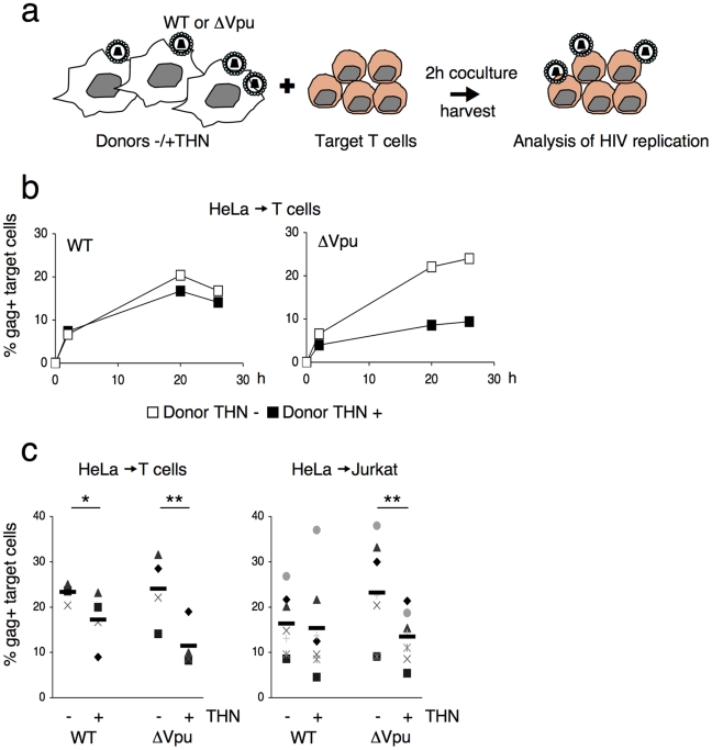
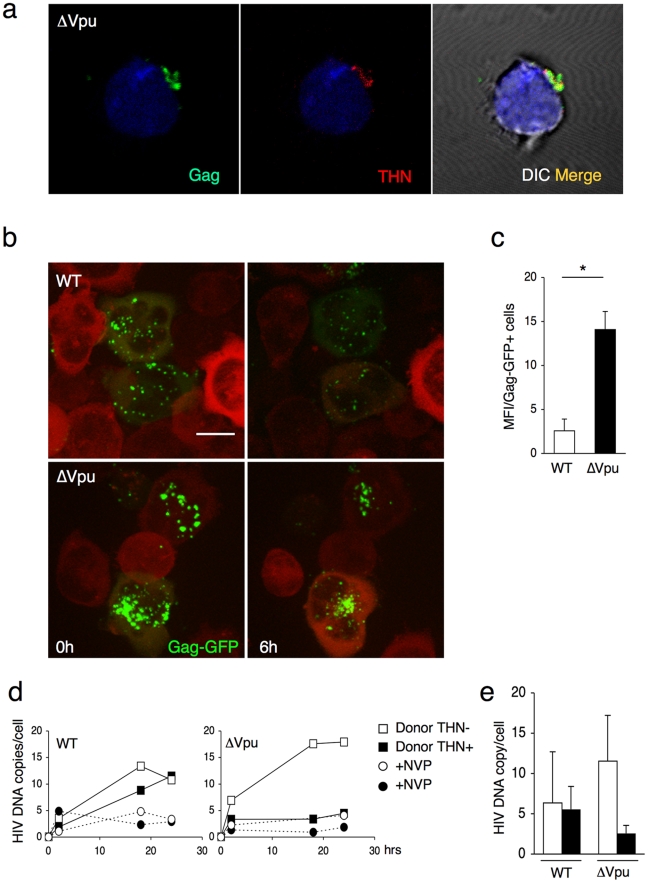
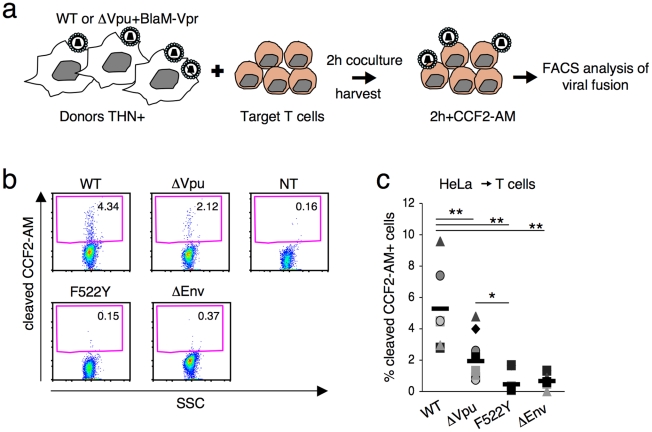
The IFN-inducible antiviral protein tetherin (or BST-2/CD317/HM1.24) impairs release of mature HIV-1 particles from infected cells. HIV-1 Vpu antagonizes the effect of tetherin. The fate of virions trapped at the cell surface remains poorly understood. Here, we asked whether tetherin impairs HIV cell-to-cell transmission, a major means of viral spread. Tetherin-positive or -negative cells, infected with wild-type or DeltaVpu HIV, were used as donor cells and cocultivated with target lymphocytes. We show that tetherin inhibits productive cell-to-cell transmission of DeltaVpu to targets and impairs that of WT HIV. Tetherin accumulates with Gag at the contact zone between infected and target cells, but does not prevent the formation of virological synapses. In the presence of tetherin, viruses are then mostly transferred to targets as abnormally large patches. These viral aggregates do not efficiently promote infection after transfer, because they accumulate at the surface of target cells and are impaired in their fusion capacities. Tetherin, by imprinting virions in donor cells, is the first example of a surface restriction factor limiting viral cell-to-cell spread.


Conflict of interest statement

The authors have declared that no competing interests exist.

Figures

Figure 1
Figure 1. Tetherin reduces HIV cell-to-cell transmission.
(a) Schematic representation of the cell-to-cell transfer assay. (b) HIV cell-to-cell transmission analyzed by flow cytometry. Hela donor cells expressing (black squares) or not expressing (white squares) tetherin (THN) were infected with WT (left panel) or ΔVpu (right panel) HIV. Cells were then cocultivated with target lymphocytes. The percentage of Gag+ cells in targets, at different time points is shown in this representative experiment. (c) Mean±sd (black line) of 4 and 7 independent experiment (20 h time point) with primary T cells (left) and Jurkat T cells (right) as targets, respectively. *p<0.05; **p<0.01 (Mann-Whitney test).
Figure 2
Figure 2. Tetherin reduces HIV cell-to-cell transmission from 293T and primary T cells.
(a) Effect of transient expression of tetherin. 293T cells donor cells were cotransfected with WT (left panel) or ΔVpu (right panel) HIV proviruses, along with a control (white squares) or a tetherin expression plasmid. Cells were then cocultivated with target Jurkat cells for 2 h. The percentage of Gag+ cells in targets, at different time points after harvesting the targets, is shown in this representative experiment. (b) Mean±sd (black line) of 6 independent experiment (20 h time point) with Jurkat T cells as targets. When stated (in 3 out of 6 experiments), cocultures were gently shaken to inhibit intercellular contacts *p<0.05; **p<0.01 (Mann-Whitney test). (c) Tetherin expression in IFN-treated 293T cells stably expressing a control shRNA (continuous line) or an shRNA targeting THN (dotted line). Grey histogram: cells not treated with IFN. Cell-to-cell transfer using infected 293T cells as donors and Jurkat as targets (right panels). Mean±sd of 3 independent experiments is depicted. (d) HIV transfer between primary T cells. Donor lymphocytes were infected with WT (white squares) or ΔVpu (black squares) HIV. The percentage of Gag+ cells among targets is shown at the indicated times points in this representative experiment. Right: mean (black line) ±sd of 4 independent experiments. Targets were treated or not with Nevirapine (NVP), a reverse transciptase inhibitor, after coculture. (e) Compilation of 3 independent experiments using MT4C5 T cells as donors and Jurkat as targets. Data are mean ±sd. b, c, e: 20 h time point; d: 48 h time point. *p<0.05; **p<0.01 (Mann-Whitney test).
Figure 3
Figure 3. Tetherin accumulates with Gag at the virological synapse.
(a) Localization of Gag and tetherin at the virological synapse. WT or ΔVpu HIV-infected MT4C5 cells were mixed with far-red-dye labeled Jurkat recipients (blue) for 1 h, and stained for HIV-1 Gag (green) and tetherin (red). Representative images are shown. (b) Quantification of infected MT4C5 cells displaying Gag clustering at the junction zone with targets. Data are mean±sd of 5 independent experiments (with a least of 650 Gag+ cells analyzed for each condition). (c) Quantification of virological synapses displaying tetherin clustering at the junction zone. Data are mean±sd of 5 independent experiments (with a least of 200 synapses analyzed for each condition). (d) Live video-microscope imaging of cell-to-cell transfer. Jurkat cells transfected with WT or ΔVpu HIV-GagGFP were mixed with actin-RFP expressing Jurkat targets and analyzed. A 3D image was acquired every 20 seconds for 2 hours. 2D maximum-projection of the images is shown Elapsed time after mixing is indicated. Corresponding sequences are supplementary Videos S1 (HIV-GagGFP WT) and S2 (HIV-GagGFP ΔVpu). *p<0.05 (Mann-Whitney test).
Figure 4
Figure 4. Distribution of transferred WT or ΔVpu viruses on target Jurkat cells.
(a) Gag signal on target cells. Jurkat cells labelled with far-red dye (blue) were harvested after 2 h of contact with WT or ΔVpu HIV-Gag-GFP transfected HeLa. Representative images are shown. In the right panel, quantification of target cells displaying large aggregates of Gag-GFP proteins after 2 h of coculture with WT or ΔVpu HIV-transfected HeLa (white) or HeLa-THN- cells (black). Data are mean±sd of 9 independent experiments (with a least of 2000 Gag+ cells analyzed for each condition). Similar results were obtained with parental WT or ΔVpu viruses lacking GFP, and staining with Gag mAbs (not shown) (b) Correlative electron microscopy analysis of Jurkat targets after coculture with ΔVpu HIV-GagGFP transfected Hela cells. Viral aggregates appearing as green spots in the IF image were visualized by SEM. Cells are stained with anti-Env MAb coupled to 20 nm-gold particles (appearing as white dots). (c) Long-lived Gag patches on target cells. Jurkat cells were harvested after 2 h of contact with ΔVpu HIV-Gag-GFP transfected HeLa and incubated at 37°C in presence of nevirapine. Representative images of gag signal at different time points after coculture are shown.*p<0.05; ***p<0.005 (Mann-Whitney test).
Figure 5
Figure 5. Tetherin promotes transfer of large viral patches and inhibits productive infection.
(a) Distribution of Gag and tetherin. Jurkat target cells were stained, after 1 h of contact with ΔVpu-infected MT4C5, for Gag (green) and tetherin (red). A representative image from 5 independent experiments is shown. (b) Live video-microscope imaging of transferred virus on target cells. Actin-RFP Jurkat target cells, incubated for 4 hours with HIV-GagGFP WT or ΔVpu transfected Hela donor cells, were harvested and plated on fibronectin-coated micro-dish. Images were taken every 5 minutes for 10 hours. Elapsed time after the beginning of acquisition is indicated. 2D maximum-projection of the images is shown. The scale bar represents 10 µm. Corresponding sequences are available as supplementary Videos S3 (HIV-GagGFP WT) and S4 (HIV-GagGFP ΔVpu). (c) Quantification of WT or ΔVpu Gag-GFP fluorescence on Jurkat target cells after 2 h incubation with Hela donor cells. The virus-associated fluorescence was measured for each condition on at least 50 individual cells. The results are expressed as the mean fluorescence intensity of viral aggregates per Gag-GFP positive cell ± sd. Results are representative of 3 independent experiments. *p<0.05 (Mann-Whitney test). (d) Time course analysis of HIV DNA synthesis in Jurkat cells by qPCR. A representative experiment is shown on the left. Target cells were treated or not with nevirapine (NVP), a reverse transcriptase inhibitor, after the coculture. (e) Mean ±sd of 3 independent experiments is shown on the right (20 h time point).
Figure 6
Figure 6. Tetherin reduces fusion after viral transfer to target cells.
(a) Schematic representation of the viral fusion assay after cell-to-cell transfer. (b) HIV fusion analyzed by cytometry. Jurkat T cells were cocultivated with donor Hela cells for 2 h, harvested and incubated at room temperature for 2 h with CCF2-AM. Viral fusion was evaluated by measuring the percentage of cells positive for cleaved CCF2-AM. A representative experiment is shown. (c) The percentage of cleaved-CCF2-AM+ cells in 7 independent experiments is shown along with the mean value (black line). *p<0.05; **p<0.01 (Mann-Whitney test).

Similar articles

Cited by

References

    1. Sattentau Q. Avoiding the void: cell-to-cell spread of human viruses. Nat Rev Microbiol. 2008;6:815–826. - PubMed
    1. Chen P, Hubner W, Spinelli MA, Chen BK. Predominant mode of human immunodeficiency virus transfer between T cells is mediated by sustained Env-dependent neutralization-resistant virological synapses. J Virol. 2007;81:12582–12595. - PMC - PubMed
    1. Rudnicka D, Feldmann J, Porrot F, Wietgrefe S, Guadagnini S, et al. Simultaneous cell-to-cell transmission of human immunodeficiency virus to multiple targets through polysynapses. J Virol. 2009;83:6234–6246. - PMC - PubMed
    1. Sherer NM, Lehmann MJ, Jimenez-Soto LF, Horensavitz C, Pypaert M, et al. Retroviruses can establish filopodial bridges for efficient cell-to-cell transmission. Nat Cell Biol. 2007;9:310–315. - PMC - PubMed
    1. Sowinski S, Jolly C, Berninghausen O, Purbhoo MA, Chauveau A, et al. Membrane nanotubes physically connect T cells over long distances presenting a novel route for HIV-1 transmission. Nat Cell Biol. 2008;10:211–219. - PubMed

Publication types

MeSH terms

Substances