. 2010 Jun 24;6(6):e1000965.
doi: 10.1371/journal.ppat.1000965.

Human cytomegalovirus UL29/28 protein interacts with components of the NuRD complex which promote accumulation of immediate-early RNA

Affiliations

Human cytomegalovirus UL29/28 protein interacts with components of the NuRD complex which promote accumulation of immediate-early RNA

Scott S Terhune et al. PLoS Pathog. .

Abstract

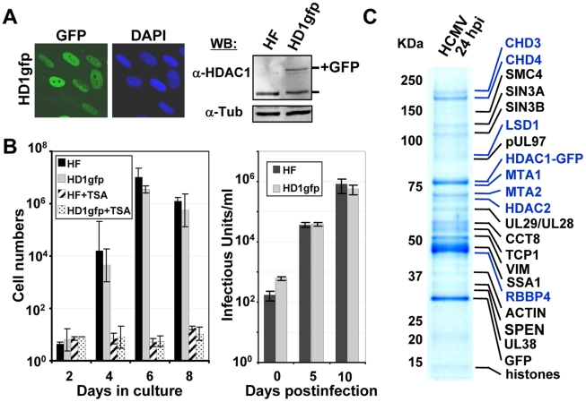
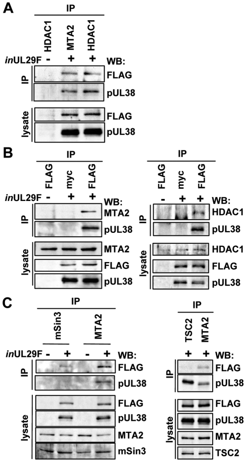
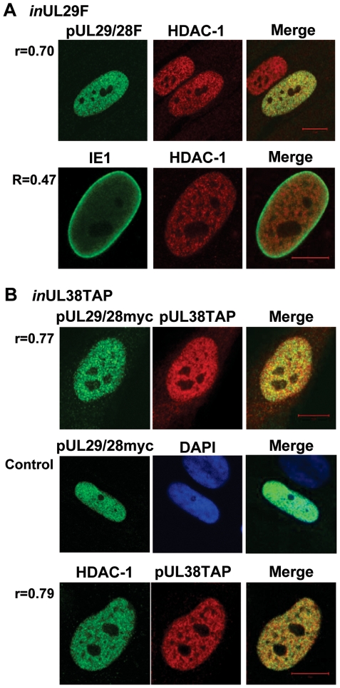
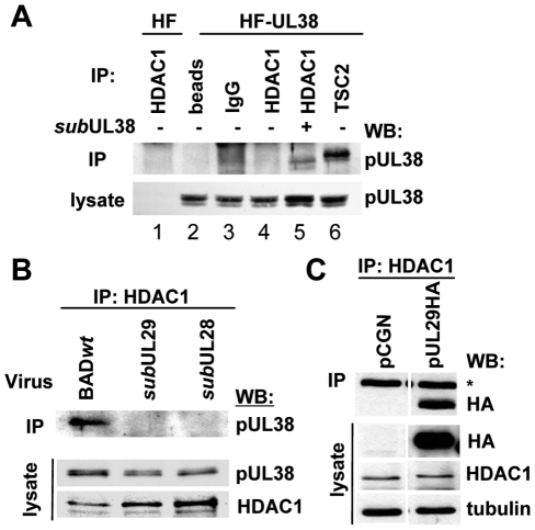
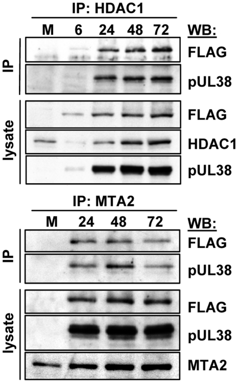
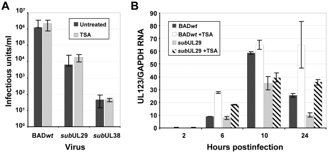
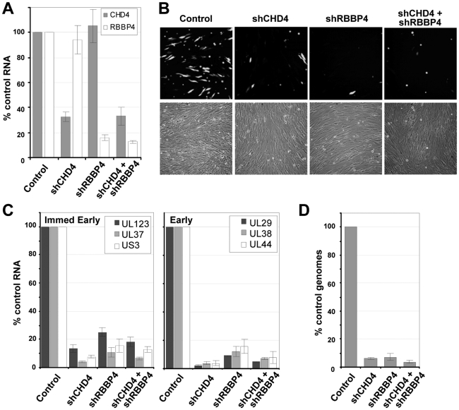
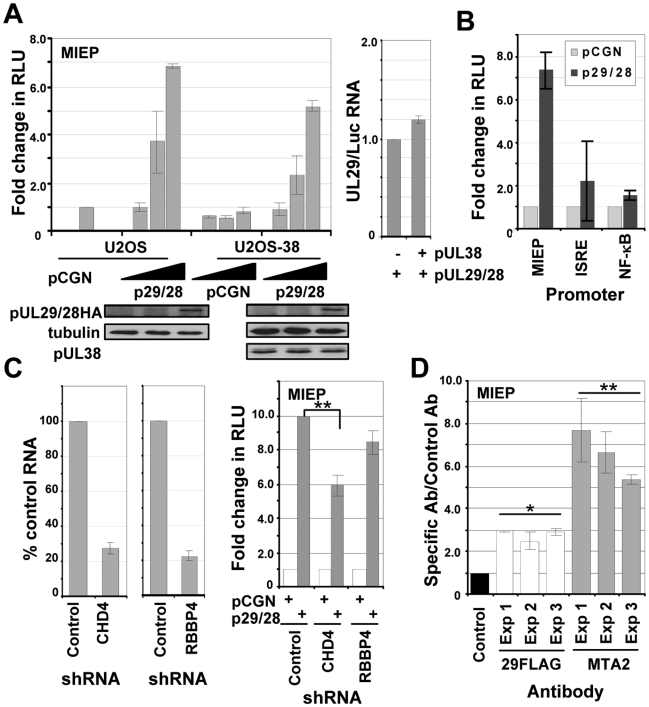
Histone deacetylation plays a pivotal role in regulating human cytomegalovirus gene expression. In this report, we have identified candidate HDAC1-interacting proteins in the context of infection by using a method for rapid immunoisolation of an epitope-tagged protein coupled with mass spectrometry. Putative interactors included multiple human cytomegalovirus-coded proteins. In particular, the interaction of pUL38 and pUL29/28 with HDAC1 was confirmed by reciprocal immunoprecipitations. HDAC1 is present in numerous protein complexes, including the HDAC1-containing nucleosome remodeling and deacetylase protein complex, NuRD. pUL38 and pUL29/28 associated with the MTA2 component of NuRD, and shRNA-mediated knockdown of the RBBP4 and CHD4 constituents of NuRD inhibited HCMV immediate-early RNA and viral DNA accumulation; together this argues that multiple components of the NuRD complex are needed for efficient HCMV replication. Consistent with a positive acting role for the NuRD elements during viral replication, the growth of pUL29/28- or pUL38-deficient viruses could not be rescued by treating infected cells with the deacetylase inhibitor, trichostatin A. Transient expression of pUL29/28 enhanced activity of the HCMV major immediate-early promoter in a reporter assay, regardless of pUL38 expression. Importantly, induction of the major immediate-early reporter activity by pUL29/28 required functional NuRD components, consistent with the inhibition of immediate-early RNA accumulation within infected cells after knockdown of RBBP4 and CHD4. We propose that pUL29/28 modifies the NuRD complex to stimulate the accumulation of immediate-early RNAs.


Conflict of interest statement

The authors have declared that no competing interests exist.

Figures

Figure 1
Figure 1. Identification of HDAC1-interacting proteins during HCMV infection.
(A) Expression of HDAC1 fused in-frame with the enhanced green fluorescent protein (GFP) in life-extended human fibroblasts. Left panel: The fusion protein was monitored by fluorescence (green), and cells were co-stained with the DNA stain, DAPI (blue). Right panel: Western blot analysis using whole cell lysates from parental (HF) or HDAC1-GFP (HD1gfp) cells and an antibody specific to HDAC1. Anti-tubulin was used as a loading control. (B) HD1gfp cells support efficient HCMV replication. Left panel: Parental (HF) and HD1gfp cell growth were monitored over time and in the presence of 500 nM trichostatin A (TSA), an HDAC inhibitor. Right panel: Replication of wild-type HCMV was quantified by infecting at a multiplicity of 0.01 pfu/cell and determining viral titers from culture supernatants. Data is from duplicate experiments. (C) HDAC1-interacting proteins. HD1gfp cells were infected at a multiplicity of 3 pfu/cell and cell lysates were collected at 24 hpi. HDAC1-GFP interacting proteins were isolated as described in the materials and methods and separated by SDS-PAGE. Gels were stained by Coomassie blue and proteins identified by mass spectrometry. Protein names highlighted in blue represent members of the NuRD complex.
Figure 2
Figure 2. HCMV pUL29/28 and pUL38 specifically bind to constituents of the NuRD complex.
Fibroblasts were infected at a multiplicity of 3 pfu/cell using BADinUL29F (inUL29F) virus, and whole cell lysates were prepared 24 h later. (A) HDAC1 and MTA2 coprecipitate with pUL29/28 and pUL38. Immunoprecipitations (IP) were performed using antibodies to MTA2 or endogenous HDAC1. Western blot (WB) analysis employed an anti-FLAG (pUL29/28) or pUL38 antibodies, and included analysis of lysates as a control. An uninfected control sample was also included. (B) pUL29/28 coprecipitates HDAC1, MTA2 and pUL38. Left panel: The immunoprecipitation was carried out using an anti-FLAG antibody (pUL29/28) or an isotype-specific anti-myc control antibody followed by Western blot analysis using antibodies to pUL38 and MTA2. Right panel: The experiment was repeated using antibody to HDAC1. (C) pUL29/28 interacts with the NuRD and Sin3 complexes. Left panel: Immunoprecipitation from whole cell lysates from uninfected or BADinUL29F (inUL29F) infected cells using antibodies to mSin3A and MTA2. Western blot analysis with antibodies against FLAG (pUL29/28), pUL38, MTA2 or mSin3A. Right panel: No evidence for interaction of pUL29/28 with tuberous sclerosis protein 2 (TSC2). Immunoprecipitations used antibodies against TSC2 and MTA2 and Western blots were performed with antibodies to FLAG (pUL29/28).
Figure 3
Figure 3. Colocalization of pUL29/28 with cellular HDAC1 and HCMV pUL38.
Colocalization is demonstrated within the merged images and quantified using Pearson's correlation (r). (A) BADinUL29F-infected cells (inUL29F). Cells were infected at a multiplicity of 0.5 infectious unit/cell, fixed at 24 hpi, and processed for immunofluorescence. Top row shows colocalization of pUL29/28 using an anti-FLAG antibody (green) and of HDAC1 (red). Bottom row demonstrates localization of IE1 (green) and HDAC1 (red) at 24 hpi. (B) BADinUL38TAP-infected cells (inUL38TAP). Cells were infected using the pUL38TAP expressing virus and fixed at 24 hpi. Top row shows expression of myc-tagged pUL29/28 using an antibody to the myc epitope (green) and of pUL38 using an antibody to protein A, detecting the TAP sequence (red). Middle row demonstrates the specific detection of pUL29/28myc (green) in the presence of uninfected cells marked by DAPI (blue). Bottom row shows colocalization of HDAC1 (green) and pUL38 (red). Pearson's correlation (r) for colocalization of fluorescent signals was determined for indicated images.
Figure 4
Figure 4. HCMV pUL29/28 is necessary for the HDAC1 interaction with pUL38.
(A) The interaction between pUL38 and HDAC1 requires additional factors present during viral infection. Immunoprecipitation (IP) from whole cell lysates isolated from either uninfected control cells (HF) or pUL38-expressing cells (HF-UL38) using antibodies against HDAC1 (lanes 1, 4) or TSC2 (lane 6). Immunoprecipitation of HDAC1 (lane 5) was also completed upon infection of HF-UL38 cells with a pUL38-deficient virus, BADsubUL38 (subUL38) at 24 hpi. Binding to pUL38 was detected by Western blot (WB) analysis with an anti-pUL38 antibody. Beads without antibody and normal IgG were used as specificity controls (lane 2, 3). (B) pUL38 binding to HDAC1 requires pUL29/28. Cells were infected at 3 pfu/cell using wild-type (BADwt), BADsubUL29 (subUL29) or BADsubUL28 (subUL28) mutant viruses and whole cell lysates collected at 24 hpi. Interactions were detected by immunoprecipitation of HDAC1 and Western blot to pUL38. Lysate controls demonstrate expression of pUL38 in all samples. (C) pUL29 binds HDAC1 in the absence of infection. Expression vectors pCGN or pCGN-pUL29HA (pUL29HA) were transfected into U-2 OS cells and whole cell lysates were collected 48 h later. Binding was demonstrated by immunoprecipitation of HDAC1 and Western blot to HA. Lysate controls show expression of pUL29HA, HDAC1 and tubulin, and an asterisks indicates heavy chain of IgG.
Figure 5
Figure 5. Accumulation of protein complexes during infection with BADinUL29F.
Fibroblasts were infected at a multiplicity of 3.0 pfu/cell and harvested at the indicated times. NuRD complexes were isolated by immunoprecipitation (IP) using an anti-HDAC1 antibody (top panels) or an anti-MTA2 antibody (bottom panels) and interacting proteins determined by Western blot (WB) assay using antibodies to pUL38, HDAC1, MTA2 and the FLAG epitope in pUL29/28.
Figure 6
Figure 6. Inhibition of deacetylase activity fails to rescue the replication of pUL29/28 or pUL38-deficient viruses.
(A) Trichostatin A does not rescue pUL29/28 or pUL38-deficient viruses. Fibroblasts were infected at a multiplicity of 0.1 pfu/cell using wild-type (BADwt) or inoculum containing equivalent numbers of viral genomes for BADsubUL29 (subUL29) or BADsubUL38 (subUL38) viruses. Cultures were treated with 300 nM TSA for the duration of the experiment and viral titers from culture supernatants were determined at 10 dpi. Data is from duplicate experiments. (B) BADsubUL29 (subUL29) virus is deficient in immediate-early gene expression irrespective of treatment with TSA. Cells pretreated with 500 nM of TSA were infected using 0.1 pfu/cell of wild-type (BADwt) or BADsubUL29 (subUL29) virus as described above, and total RNA was harvested at the indicated times. Expression of IE1 RNA was determined by qRT-PCR using cDNA produced from DNase-treated RNA and random hexamers. Quantitation was completed with SYBR green and primers specific to exon 4 of IE1. Samples were normalized to cellular GAPDH RNA levels.
Figure 7
Figure 7. Reduced expression of components of the NuRD complex inhibits HCMV replication.
(A) Disruption of NuRD complex in primary fibroblasts. Short hairpin RNA (shRNA) sequences to a scrambled control, CHD4, RBBP4 or both CHD4 and RBBP4 were delivered to fibroblasts using lentivirus vectors and expressing cells were isolated by puromycin resistance. Expression of CHD4 and RBBP4 was quantified by qRT-PCR using two separate sets of gene-specific primers and total cellular RNA. The data was normalized to GAPDH RNA levels and includes the average percent reduction for both primer sets to each gene. (B) shRNA-expressing cells were infected at a multiplicity of 0.25 pfu/cell with a BADwt derivative expressing GFP. Images of infected cells were captured at 96 hpi. (C) Expression of immediate-early and early RNAs at 10 hpi was determined by qRT-PCR using cDNA produced from DNase-treated RNA and random hexamers. Quantitation was completed with SYBR green and sequence-specific primers as indicated. Expression was normalized to cellular GAPDH RNA levels. (D) HCMV genome replication was quantified by qPCR at 96 hpi. qPCR data was normalized using primers to β-actin. Data is derived from replicate experiments.
Figure 8
Figure 8. pUL29/28 specifically activates the MIE promoter irrespective of HCMV pUL38 expression.
(A) pUL29/28 activates the MIEP. Upper left panel: U2OS cells that stably maintained the empty pLXSN vector (U2OS) or pLXSN expressing HCMV pUL38 (USOS-38) were transfected with 50 ng of pGL3-MIEP reporter plasmid and 10, 100, or 500 ng of pCGN empty vector or pCGN-pUL29/28 effector plasmid. Luciferase activity was assayed 48 h posttransfection using equal protein amounts within each lysate and normalized to luciferase activity from empty vector. Lower panels: The levels of pUL29/28HA and pUL38 expression were assayed by Western blot analysis using the same lysates and antibody to HA, pUL38 or tubulin. Right panel: UL29 and luciferase RNA expression was determined by qRT-PCR from U2OS as compared to U2OS-38 cells. (B) pUL29/28 exhibits promoter-specific effects. Luciferase assays were completed using 500 ng of pCGN or pCGN-pUL29/28 and MIEP, ISRE and NF-κB promoter reporter constructs. The relative luciferase activity was determined as described above. (C) NuRD is required for optimal expression of an MIEP reporter. Left panel: Disruption of the NuRD complex in U-2 OS cells. Short hairpin RNA (shRNA) sequences to a scrambled control, CHD4 or RBBP4 were delivered to U-2 OS cells using lentiviruses and expressing cells were isolated by puromycin resistance. Expression of CHD4 and RBBP4 was quantified by qRT-PCR. The data was normalized to GAPDH RNA levels and includes the percent reduction for each gene relative to control. Right panel: Luciferase assays were completed using either empty pCGN or pCGN-pUL29/28 and MIEP reporter. The relative luciferase activity was determined as described above and the data is derived from replicate experiments (**p<0.03). (D) pUL29/28 and MTA2 interact with the MIE promoter during infection. Cells were infected using BADinUL29F at a multiplicity of 3 pfu/cell and harvested at 48 hpi. Antibodies to the FLAG epitope in pUL29/28FLAG, cellular MTA2 and a control antibody to HCMV pUS24 were used to immunoprecipitate chromatin-DNA complexes, and isolated DNA was quantified by qPCR and primers to the HCMV MIE promoter. The results are plotted as the ratio of viral DNA immunoprecipitated to input DNA for each antibody relative to that of the control antibody. The data present the average of two separate qPCR quantifications from three separate experiments. Significance of changes for the pooled sets of samples from three experiments were analyzed by student's t-test, and the p value is indicated by an asterisk (*p<0.007,**p<0.03).

Similar articles

Cited by

References

    1. Mocarski E, Shenk T, Pass RF. Fields Virology: Lippincott Williams & Wilkins; 2007. Cytomegaloviruses. pp. 2702–2772.
    1. Streblow DN, Dumortier J, Moses AV, Orloff SL, Nelson JA. Mechanisms of cytomegalovirus-accelerated vascular disease: induction of paracrine factors that promote angiogenesis and wound healing. Curr Top Microbiol Immunol. 2008;325:397–415. - PMC - PubMed
    1. Waller EC, Day E, Sissons JG, Wills MR. Dynamics of T cell memory in human cytomegalovirus infection. Med Microbiol Immunol. 2008;197:83–96. - PubMed
    1. Cinatl J, Jr, Nevels M, Paulus C, Michaelis M. Activation of telomerase in glioma cells by human cytomegalovirus: another piece of the puzzle. J Natl Cancer Inst. 2009;101:441–443. - PubMed
    1. Terhune SS, Schroer J, Shenk T. RNAs are packaged into human cytomegalovirus virions in proportion to their intracellular concentration. J Virol. 2004;78:10390–10398. - PMC - PubMed

Publication types

MeSH terms