. 2010 Jun 24;5(6):e10964.
doi: 10.1371/journal.pone.0010964.

Human endometrial side population cells exhibit genotypic, phenotypic and functional features of somatic stem cells

Affiliations

Human endometrial side population cells exhibit genotypic, phenotypic and functional features of somatic stem cells

Irene Cervelló et al. PLoS One. .

Abstract

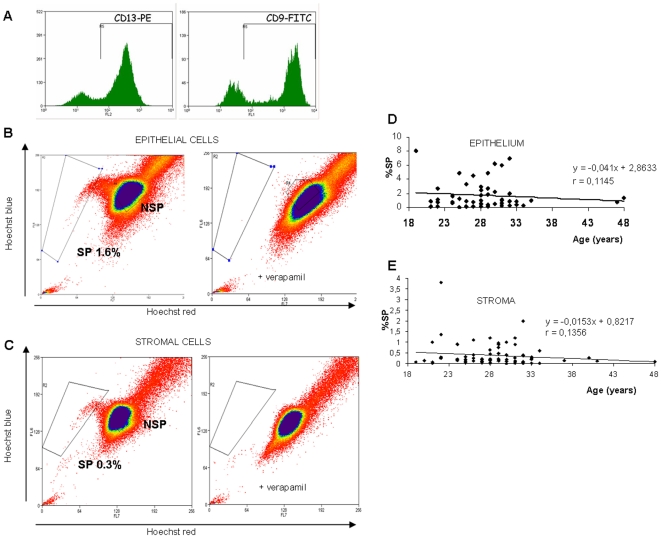
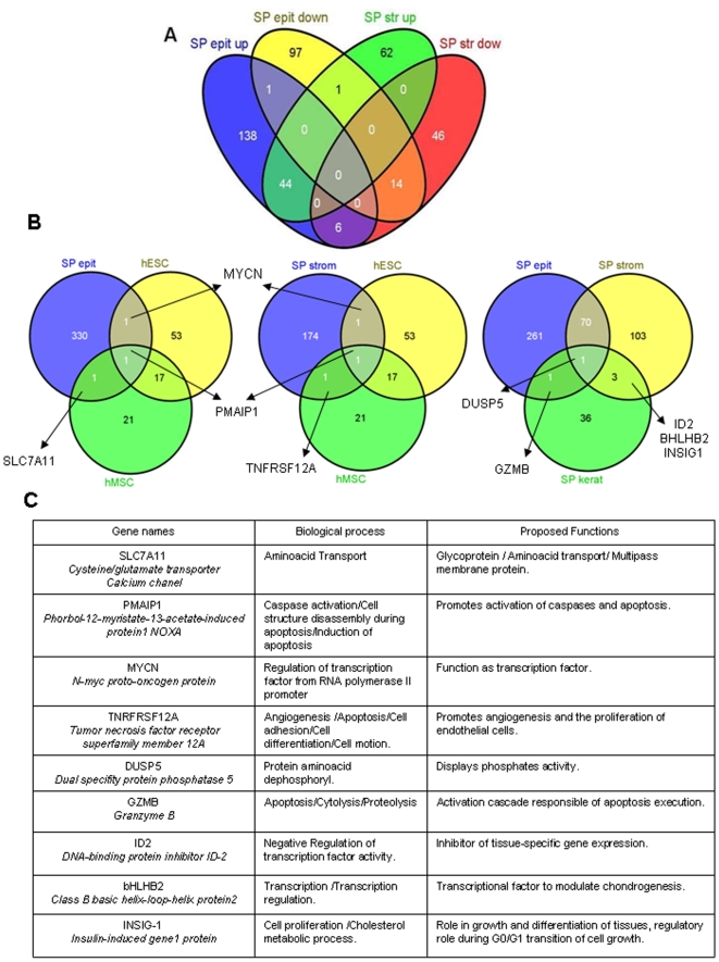
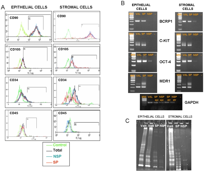
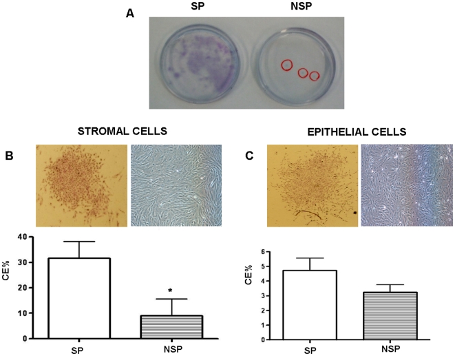
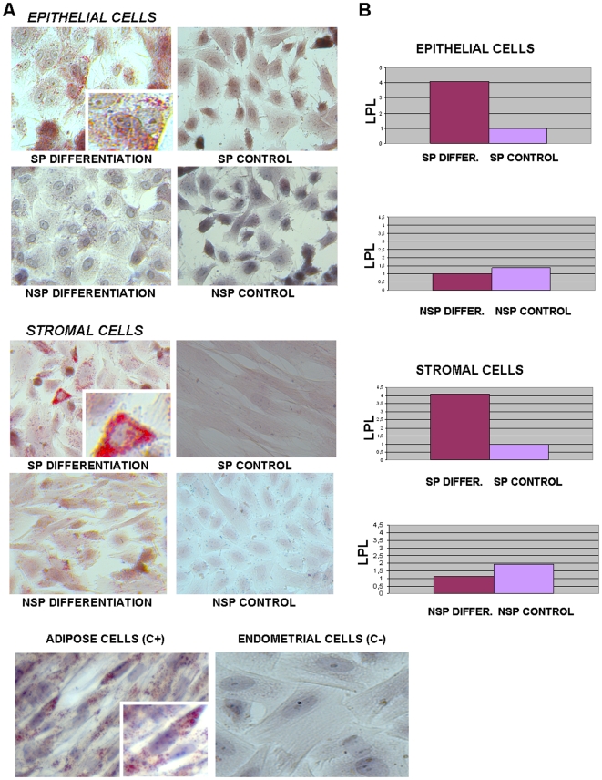
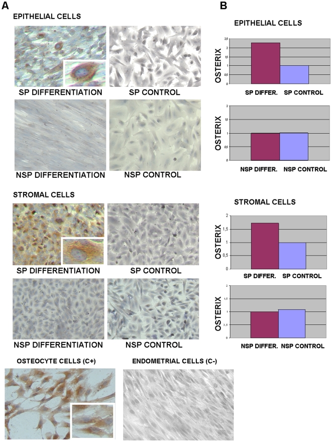
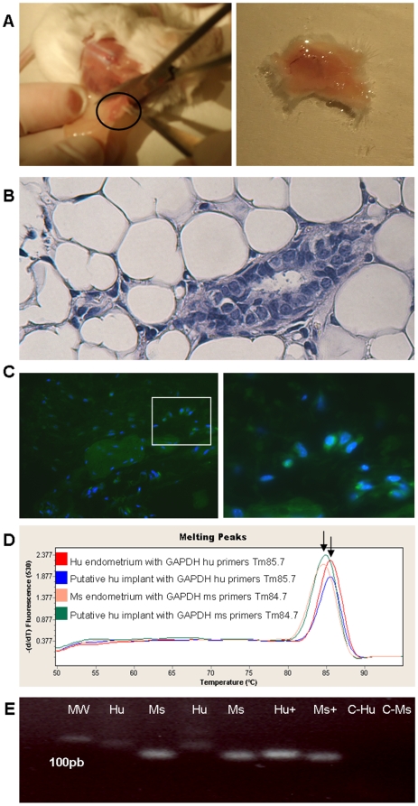
During reproductive life, the human endometrium undergoes around 480 cycles of growth, breakdown and regeneration should pregnancy not be achieved. This outstanding regenerative capacity is the basis for women's cycling and its dysfunction may be involved in the etiology of pathological disorders. Therefore, the human endometrial tissue must rely on a remarkable endometrial somatic stem cells (SSC) population. Here we explore the hypothesis that human endometrial side population (SP) cells correspond to somatic stem cells. We isolated, identified and characterized the SP corresponding to the stromal and epithelial compartments using endometrial SP genes signature, immunophenotyping and characteristic telomerase pattern. We analyzed the clonogenic activity of SP cells under hypoxic conditions and the differentiation capacity in vitro to adipogenic and osteogenic lineages. Finally, we demonstrated the functional capability of endometrial SP to develop human endometrium after subcutaneous injection in NOD-SCID mice. Briefly, SP cells of human endometrium from epithelial and stromal compartments display genotypic, phenotypic and functional features of SSC.


Conflict of interest statement

Competing Interests: The authors have declared that no competing interests exist.

Figures

Figure 1
Figure 1. Isolation of purified epithelial and stromal SP cells.
(A) Purity of the stromal and epithelial cell fractions was assessed by immunocytochemistry and flow-cytometry, 79.6% of the cells were CD13+ (stroma), and 75.8% were CD9+ (epithelium). (B & C) Distribution of the SP and NSP populations of living cells isolated from the epithelium (B), and stroma (C) of the human endometrium. (Right) coadittion of 100 µm verapamil resulted in the disappearance of the SP fraction. (D & E) Correlation tables showing the percentage of SP in the epithelium (D) and stroma (E) through womeńs reproductive life.
Figure 2
Figure 2. Endometrial SP Gene Signature.
(A) The Venn diagram represents the numbers of shared genes up- and down-regulated in the epithelial and stromal SP of the human endometrium (see Table S1 for details). (B) Venn diagrams representing: (left) the number of shared genes expressed by epithelial SP, human embryonic stem cells (hESC), and human mesenchymal stem cells (hMSC). (Center) shared genes expressed by the stromal SP, hESC and hMSC. (Right) the Venn diagram represents the number of shared genes expressed by epithelial SP, stromal SP and SP from the keratinocytes (see Table S3 for details). (C) Table describing the biological process and proposed functions of the consensus genes obtained in the previous Venn diagrams.
Figure 3
Figure 3. Phenotype of epithelial and stromal SP and telomerase activity.
(A) Sorted SP, NSP and total endometrial fraction for epithelium and stroma were fixed and positive cells were quantified by FACS for mesenchymal markers (CD90 & CD105) and hematopoietic markers (CD34 & CD45). (B) Sorted SP versus NSP cells were analyzed by PCR and nested-PCR showing the bands corresponding to typical undifferentiated markers c-KIT and OCT-4 as well as Side Population/ABC transporter markers like BCRP1 and MDR1. VAL corresponds to the human embryonic stem cell line VAL-4 obtained from the Spanish Stem Cell Bank (http://www.isciii.es/htdocs/terapia/terapia_lineas.jsp). (C)Telomerase activity of sorted SP and NSP from the stromal and epithelial compartments are presented in this figure having as a positive control VAL-4. In both compartments SP depicted an intermediate telomerase pattern compared to hESC and NSP.
Figure 4
Figure 4. Cloning efficiency under hypoxic conditions.
(A) Human endometrial cell clones under hypoxic conditions. Typical colonies formed by sorted SP (left) and NSP (right) human endometrial stromal cells were seeded at clonal densities and cultured for 15 days under hypoxic conditions. (B) Colony morphology (upper panel) and cloning efficiency (%) of sorted stromal SP that was significantly greater than NSP cultured in hypoxic conditions (p = 0,018). (C) Colony morphology (upper panel) and cloning efficiency (%) of sorted epithelial SP versus NSP cultured in hypoxic conditions. (Data are means ± SEM)
Figure 5
Figure 5. Adipogenic differentiation in vitro of epithelial and stromal SP.
(A) Induction of adypocite differentiation of epithelial and stromal SP, but not NSP, determined by Oil Red O staining of lipids droplets. Staining with Oil Red O revealed the presence of lipids vacuoles in the sorted endometrial SP cells versus NSP and their controls cultured without differentiation media.Positive control are adipose cells and negative control are non-sorted endometrial cells (lower panel). (B) Induction of adypocite differentiation of epithelial and stromal SP, but not NSP, determined by real-time PCR of adipocyte-specific gene lipoprotein lipase (LPL) demonstrated differential gene expression of SP versus NSP and their controls.
Figure 6
Figure 6. Osteogenic differentiation in vitro of epithelial and stromal SP.
(A) Induction of osteogenic differentiation of epithelial and stromal SP, but not NSP, determined by immunocytochemistry with bone sialoprotein. Staining with a protein highly specific for bone revealed the presence of osteoblast in the sorted endometrial SP cells versus NSP and their controls cultured without differentiation media. Positive controls are osteocyte cells from explants and negative control are non-sorted endometrial cells (lower panel). (B) Induction of osteocyte differentiation of epithelial and stromal SP, but not NSP, determined by real-time PCR of osterix and osteonectin demonstrated differential gene expression of SP versus NSP and their controls.
Figure 7
Figure 7. Reconstruction of human endometrial-like tissue from SP in NOD-SCID mice.
(A) Macroscopic finding of the injected site of sorted SP cells sixty days after transplantation in NOD-SCID mice showing putative endometrial like implant in subcutaneous back tissue. (B) H&E staining of putative human endometrial glands surrounded by mice adipose tissue in the injection site. (C) Left, immunohistochemical analysis for Human Progesterone Receptor (Hu-PR) in endometrial like structures in mice subcutaneous tissue obtained after injection of SP cells from human stroma (40X). Right, Detail of green fluorescent signal due to Hu-PR co-localized with DAPI in a putative human gland (High magnification: 63X-oil immersion microscopy). Negative controls with deletion of the antibody and using mice tissue were satisfactory. (D) Real Time diagram showing melting peaks. The difference between both Melting Temperatures (Tm) was an indicator of the two existing amplicons (specific nucleotide sequence) corresponding to the two different species (human and mouse). (E) m-RNA expression of human GAPDH (79 bp) and mouse GAPDH (75 bp) in reconstructed human tissue determined by real-time PCR. MW (lane 1): molecular weight pattern. Hu (lane 2 and 4): laser microdissected glandular tissue extracted from xenotransplanted mice hybridized with human primers. Ms (lanes 3 and 5): laser microdissected glandular tissue extracted from xenotransplanted mice hybridized with mice primers. Hu+ (lane 6): human endometrial tissue hybridized with human primers. Ms+ (lane 7): mouse endometrium hybridized with mice primers. C-Hu (lane 8): human endometrial tissue hybridized with mice primers. C-Ms (lane 9): mouse endometrium hybridized with human primers. All bands were sequenced confirming the human tissue presence in the laser microdissected glands extracted from mouse adipose tissue.

Similar articles

Cited by

References

    1. Prianishnikov VA. On the concept of stem cell and a model of functional-morphological structure of the endometrium. Contraception. 1978;18(3):213–23. - PubMed
    1. Padykula AH. Regeneration in the primate uterus: the role of stem cells. Ann N Y Acad Sci. 1991;622:47–56. - PubMed
    1. Reynolds BA, Weiss S. Generation of neurons and astrocytes from isolated cells of the adult mammalian central nervous system. Science. 1992;255(5052):1646. - PubMed
    1. Gage FH. Structural plasticity: cause, result, or correlate of depression. Biol Psychiatry. 2000;48(8):713–4. - PubMed
    1. Bjerknes M, Cheng H. Clonal analysis of mouse intestinal epithelial progenitors. Gastroenterology. 1999;116:208–10. - PubMed

Publication types