. 2010 Jun 24;5(6):e11301.
doi: 10.1371/journal.pone.0011301.

The terminal immunoglobulin-like repeats of LigA and LigB of Leptospira enhance their binding to gelatin binding domain of fibronectin and host cells

Affiliations

The terminal immunoglobulin-like repeats of LigA and LigB of Leptospira enhance their binding to gelatin binding domain of fibronectin and host cells

Yi-Pin Lin et al. PLoS One. .

Abstract

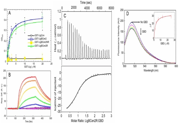
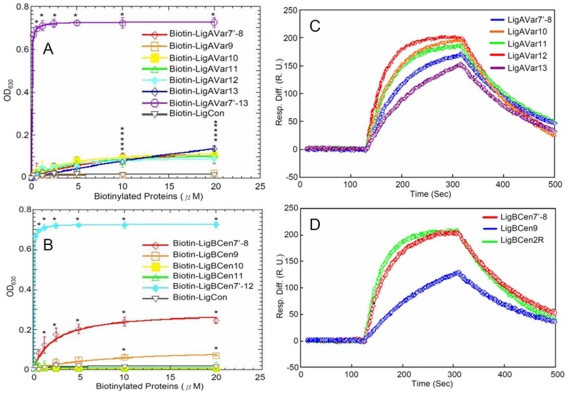
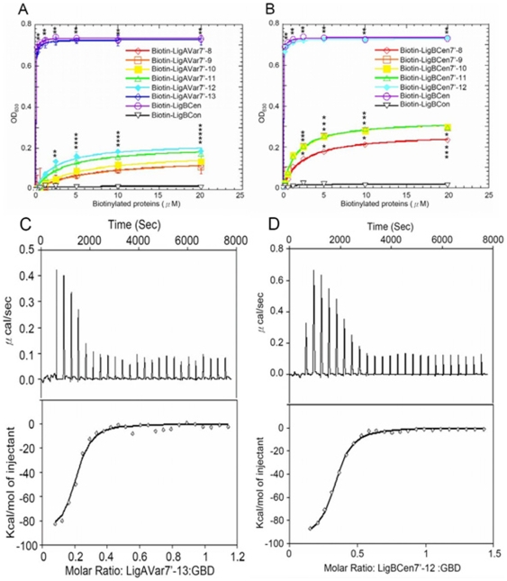
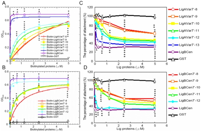
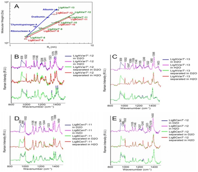
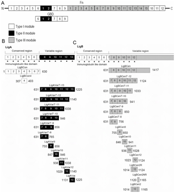
Leptospira spp. are pathogenic spirochetes that cause the zoonotic disease leptospirosis. Leptospiral immunoglobulin (Ig)-like protein B (LigB) contributes to the binding of Leptospira to extracellular matrix proteins such as fibronectin, fibrinogen, laminin, elastin, tropoelastin and collagen. A high-affinity Fn-binding region of LigB has been localized to LigBCen2, which contains the partial 11th and full 12th Ig-like repeats (LigBCen2R) and 47 amino acids of the non-repeat region (LigBCen2NR) of LigB. In this study, the gelatin binding domain of fibronectin was shown to interact with LigBCen2R (K(D) = 1.91+/-0.40 microM). Not only LigBCen2R but also other Ig-like domains of Lig proteins including LigAVar7'-8, LigAVar10, LigAVar11, LigAVar12, LigAVar13, LigBCen7'-8, and LigBCen9 bind to GBD. Interestingly, a large gain in affinity was achieved through an avidity effect, with the terminal domains, 13th (LigA) or 12th (LigB) Ig-like repeat of Lig protein (LigAVar7'-13 and LigBCen7'-12) enhancing binding affinity approximately 51 and 28 fold, respectively, compared to recombinant proteins without this terminal repeat. In addition, the inhibited effect on MDCKs cells can also be promoted by Lig proteins with terminal domains, but these two domains are not required for gelatin binding domain binding and cell adhesion. Interestingly, Lig proteins with the terminal domains could form compact structures with a round shape mediated by multidomain interaction. This is the first report about the interaction of gelatin binding domain of Fn and Lig proteins and provides an example of Lig-gelatin binding domain binding mediating bacterial-host interaction.


Conflict of interest statement

Competing Interests: The authors have declared that no competing interests exist.

Figures

Figure 1
Figure 1. Determination of binding constant, kinetics and thermodynamic parameters of the LigBCen2R/GBD interaction by ELISA, SPR, and ITC.
(A) Binding of serial concentrations of LigBCen2NR to immobilized GBD by ELISA. Serial concentrations of GST-LigBCen2R, GST-LigBCen2NR, GST-LigBCen2 (positive control), or GST-LigCon (negative control) were added to 1 µM of GBD or BSA coated wells (negative control, data not shown). (B) SPR analysis of LigBCen2R interacting with GBD. 1.5 µM of Recombinant Histidine-tagged LigBCen2R was immobilized on the surface of a Ni-NTA chip. GBD in Tris Buffer containing 100 µM CaCl2 at pH 7.5 flowed through the chip and the concentrations of GBD ranged from 40 to 0.625 µM (from top to bottom). The KD, kon, and koff were obtained from the average of duplicate experiments shown in Table 1. (C) Determination of the binding affinity by ITC. The cell contained 1 ml of GBD and the syringe contained 250 µl of LigBCen2R (upper panel). Heat differences obtained from 25 injections of LigBCen2R; (lower panel). Integrated curve with experimental data (⋄) and the best fit (-). The thermodynamic parameters are shown as the average of duplicate experiments (KD = 1.88±0.09 µM, ΔH = −29.25±3.58 kcal mol-1, TΔS = −21.47±3.61 kcal mol-1 K-1, n = 0.995±0.01). (D) Fluorescence spectrum of Alexa-488 labeled LigBCen2RW1073C in the presence and absence of GBD. One µM of Alexa-488 labeled LigBCen2RW1073C in Tris buffer was excited at 485 nm. Aliquots of GBD from respective stock solutions were added. The figure shows Alexa488 fluorescence in the presence of 0, 1.62, 3.12, 6.25, 12.5, 25 µM of GBD (Inner plot). The determination of KD of Alexa488 labeled LigBCen2RW1073C and GBD by monitoring the quenching fluorescence intensity of Alexa488 labeled LigBCen2RW1073C titrated by GBD. The emission wavelength recorded in this figure was 513 nm, and KD was revealed by fitting the data point into the equation described in materials and methods (KD = 1.93.4 µM).
Figure 2
Figure 2. Localization of the GBD-binding domains on Lig proteins.
(A and B) Various concentrations (0, 0.3125, 0.625, 1.25, 2.5, 5, 10, 20 µM) of biotinylated LigCon (negative control), and (A) LigAVar7'-13 (positive control), LigAVar7'-8, LigAVar9, LigAVar10, LigAVar11, LigAVar12, LigAVar13, (B) LigBCen7'-12 (positive control), LigBCen7'-8, LigBCen9, LigBCen10, LigBCen11 were added to wells coated with 1 µM of GBD or BSA (negative control and data not shown) in Tris buffer. The binding of biotinylated proteins to GBD was measured by ELISA. For all experiments, each value represents the mean±SEM of three trials in triplicate samples. Statistically significant (p<0.05) differences compared to the negative control are indicated by an asterisk. (C and D) SPR analysis of Ig-like domains of Lig interacting with GBD 1.5 µM of Recombinant Histdine-tag (C) LigAVar7'-8, LigAVar10, LigAVar11, LigAVar12, LigAVar13, (D) LigBCen7'-8, LigBCen9, or LigBCen2R was immobilized on the surface of Ni-NTA chip. GBD in Tris Buffer containing 100 µM CaCl2 at pH 7.5 was flowed through the chip and the concentration of GBD ranged from 40 to 0.625 µM. Only the response sensograms of the 40 µM of GBD are shown. The KD, kon, koff were obtained from the average of duplicate experiments shown in Table 1.
Figure 3
Figure 3. Terminal Ig-like domains mainly contributes to Lig proteins binding to GBD of Fn.
(A and B) The binding of GBD to various length Lig proteins constructs performed by ELISA. Various concentrations (0, 0.3125, 0.625, 1.25, 2.5, 5, 10, 20 µM) of biotinylated LigCon (negative control), LigBCen (positive control), and (A) LigAVar7'-8, LigAVar7'-9, LigAVar7'-10, LigAVar7'-11, LigAVar7'-12, or LigAVar7'-13, (B) LigBCen7'-8, LigBCen7'-9, LigBCen7'-10, LigBCen7'-11, or LigBCen7'-12 were added to wells coated with 1 µM of GBD or BSA (negative control and data not shown) in Tris buffer. The binding of biotinylated proteins to GBD was measured by ELISA. For all experiments, each value represents the mean±SEM of three trials in triplicate samples. Statistically significant (p<0.05) differences compared to the negative control are indicated by an asterisk. The KD were obtained from the average of triplicate experiments shown in Table 2. (C and D) Representative ITC isotherms showing the binding of GBD to various length LigA or LigB constructs. The cell contained 1 ml of GBD and the syringe contained 250 µl of (A) LigAVar7'-13, and (B) LigBCen7'-12, (upper panel). Heat differences obtained from 25 injections of LigAVar7'-13 or LigBCen7'-12 (lower panel); Integrated curve with experimental data (⋄) and the best fit (-). The thermodynamic parameters are shown as the average of duplicate experiments in Table 3.
Figure 4
Figure 4. Terminal Ig-like domains mainly mediates Lig protein binding and Leptospiral adhesion on MDCK cells.
(A and B) Binding of truncated Lig proteins to MDCK cells. Various concentrations (0.08, 0.16, 0.3125, 0.625, 1.25, 2.5, 5 µM) of biotinylated LigCon (negative control), LigBCen (positive control), and (A) LigAVar7'-13, LigAVar7'-12, LigAVar7'-11, LigAVar7'-10, LigAVar7'-9, LigAVar7'-8, (B) LigBCen7'-12, LigBCen7'-11, LigBCen7'-10, LigBCen7'-9, LigBCen7'-8 were added to MDCK cells (105), and binding was measured by ELISA. (C and D) Truncated Lig proteins inhibit the binding of Leptospira to MDCK cells. MDCK cells were incubated with various concentrations (0.08, 0.16, 0.3125, 0.625, 1.25, 2.5, 5 µM) of biotinylated -LigB1706-1716, or biotin (negative control) (C) LigAVar7'-13, LigAVar7'-12, LigAVar7'-11, LigAVar7'-10, LigAVar7'-9, LigAVar7'-8, (D) LigBCen7'-12, LigBCen7'-11, LigBCen7'-10, LigBCen7'-9, LigBCen7'-8 prior to the addition of Leptospira (107). The adhesion of Leptospira to MDCK cells (105) was detected by ELISA. The reduced percentage of attachment was determined relative to the attachment of Leptospira on untreated MDCK cells. Each value represents the mean±SEM of three trials in triplicate samples. Statistically significant values (P<0.05) are indicted by *.
Figure 5
Figure 5. Terminal Ig-like domains contribute to the compact structure of Lig proteins.
(A) Dynamic light scattering of standard molecular mass markers, LigAVar7'-13, LigAVar7'-12, LigAVar7'-11, LigAVar7'-10, LigAVar7'-9, LigAVar7'-8, LigBCen7'-12, LigBCen7'-11, LigBCen7'-10, LigBCen7'-9, LigBCen7'-8. The hydrodynamic radius (Rh) is plotted as a function of the molecular weight of each individual protein on a log-log scale. The molecular weights of the standards are indicated in Materials and Methods, and the molecular weight of the truncated Lig proteins are indicated in Table 5. (B to E) Comparative Raman spectroscopy of full-length LigAVar7'-12, LigAVar7'-13, LigBCen7'-11, LigBCen7'-12 and a stoichiometic mixture of its composite domains. The Raman spectra of full length or stoichiometrically mixed separated Ig-like domains of (B) LigAVar7'-12, (C) LigAVar7'-13, (D) LigBCen7'-11, and (E) LigBCen7'-12 were recorded in D2O and H2O.
Figure 6
Figure 6. A schematic diagram showing the structure of truncated (A) Fn, (B) LigA, and (C) used in this study.

Similar articles

Cited by

References

    1. Patti JM, Allen BL, McGavin MJ, Hook M. MSCRAMM-mediated adherence of microorganisms to host tissues. Annu Rev Microbiol. 1994;48:585–617. - PubMed
    1. Potts JR, Campbell ID. Fibronectin structure and assembly. Curr opin cell Biol. 1994;6(5):648–655. - PubMed
    1. Vakonakis I, Campbell ID. Extracellular matrix: from atomic resolution to ultrastructure. Curr Opin Cell Biol. 2007;19(5):578–583. - PMC - PubMed
    1. Schwarz-Linek U, Hook M, Potts JR. The molecular basis of fibronectin-mediated bacterial adherence to host cells. Mol Microbiol. 2004;52(3):631–641. - PubMed
    1. Joh D, Speziale P, Gurusiddappa S, Manor J, Hook M. Multiple specificities of the staphylococcal and streptococcal fibronectin-binding microbial surface components recognizing adhesive matrix molecules. Eur J Biochem. 1998;258(2):897–905. - PubMed

Publication types