

Experimental human cell and tissue models of pemphigus
- PMID: 20585596
- PMCID: PMC2877615
- DOI: 10.1155/2010/143871
Experimental human cell and tissue models of pemphigus
Abstract
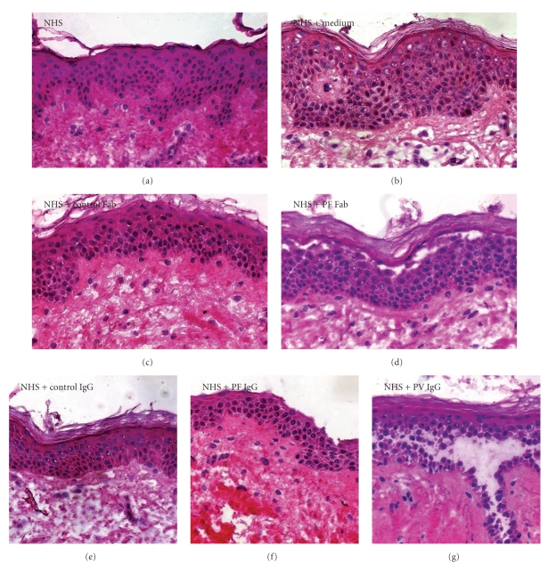
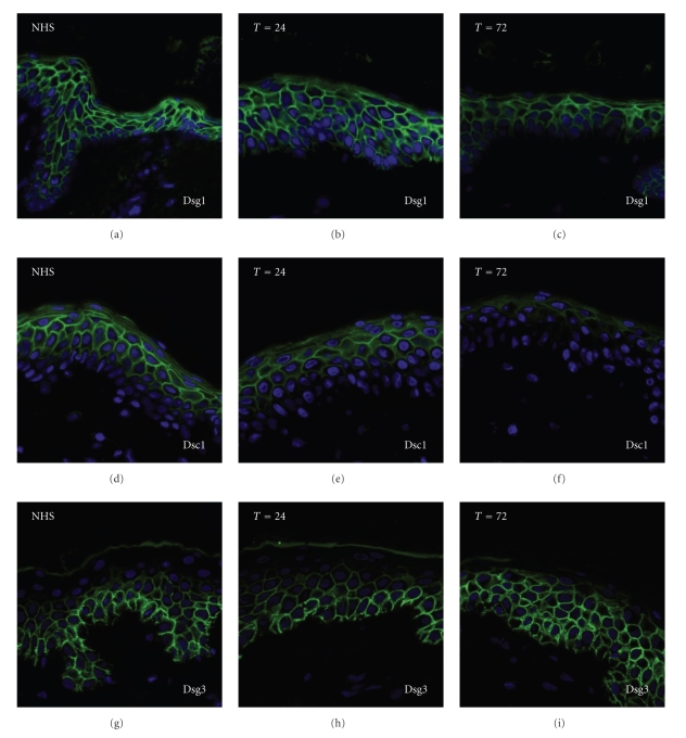
Pemphigus is a chronic mucocutaneous autoimmune bullous disease that is characterized by loss of cell-cell contact in skin and/or mucous membranes. Past research has successfully identified desmosomes as immunological targets and has demonstrated that acantholysis is initiated through direct binding of IgG. The exact mechanisms of acantholysis, however, are still missing. Experimental model systems have contributed considerably to today's knowledge and are still a favourite tool of research. In this paper we will describe to what extent human cell and tissue models represent the in vivo situation, for example, organ cultures of human skin, keratinocyte cultures, and human skin grafted on mice and, furthermore, how suitable they are to study the pathogenesis of pemphigus. Organ cultures closely mimic the architecture of the epidermis but are less suitable to answer posed biochemical questions. Cultured keratinocyte monolayers are convenient in this respect, but their desmosomal make-up in terms of adhesion molecules does not exactly reflect the in vivo situation. Reconstituted skin is a relatively new model that approaches organ culture. In models of human skin grafted on mice, acantholysis can be studied in actual human skin but now with all the advantages of an animal model.
Figures
Similar articles
-
Mechanisms Causing Acantholysis in Pemphigus-Lessons from Human Skin.Front Immunol. 2022 May 20;13:884067. doi: 10.3389/fimmu.2022.884067. eCollection 2022. Front Immunol. 2022. PMID: 35720332 Free PMC article. Review.
-
Apoptotic mechanism in pemphigus autoimmunoglobulins-induced acantholysis--possible involvement of the EGF receptor.Autoimmunity. 2006 Nov;39(7):563-75. doi: 10.1080/08916930600971836. Autoimmunity. 2006. PMID: 17101500
-
Ultrastructural studies of acantholysis induced in vivo by passive transfer of IgG from endemic pemphigus foliaceus (Fogo Selvagem).J Invest Dermatol. 1989 Oct;93(4):480-5. doi: 10.1111/1523-1747.ep12284041. J Invest Dermatol. 1989. PMID: 2778350
-
Protease inhibitors prevent plasminogen-mediated, but not pemphigus vulgaris-induced, acantholysis in human epidermis.Biol Chem. 2003 Feb;384(2):311-5. doi: 10.1515/BC.2003.035. Biol Chem. 2003. PMID: 12675525
-
Desmosomes and disease: pemphigus and bullous impetigo.Curr Opin Cell Biol. 2004 Oct;16(5):536-43. doi: 10.1016/j.ceb.2004.07.006. Curr Opin Cell Biol. 2004. PMID: 15363804 Review.
Cited by
-
Electron microscopy of desmosomal structures in the pemphigus human skin organ culture model.Front Med (Lausanne). 2022 Nov 14;9:997387. doi: 10.3389/fmed.2022.997387. eCollection 2022. Front Med (Lausanne). 2022. PMID: 36452895 Free PMC article.
-
Endocytosis of IgG, Desmoglein 1, and Plakoglobin in Pemphigus Foliaceus Patient Skin.Front Immunol. 2019 Nov 12;10:2635. doi: 10.3389/fimmu.2019.02635. eCollection 2019. Front Immunol. 2019. PMID: 31781120 Free PMC article.
-
No evidence of apoptotic cells in pemphigus acantholysis.J Invest Dermatol. 2014 Jul;134(7):2039-2041. doi: 10.1038/jid.2014.60. Epub 2014 Jan 31. J Invest Dermatol. 2014. PMID: 24487306 No abstract available.
-
The extent of desmoglein 3 depletion in pemphigus vulgaris is dependent on Ca(2+)-induced differentiation: a role in suprabasal epidermal skin splitting?Am J Pathol. 2011 Oct;179(4):1905-16. doi: 10.1016/j.ajpath.2011.06.043. Epub 2011 Aug 22. Am J Pathol. 2011. PMID: 21864491 Free PMC article.
-
Mechanisms Causing Acantholysis in Pemphigus-Lessons from Human Skin.Front Immunol. 2022 May 20;13:884067. doi: 10.3389/fimmu.2022.884067. eCollection 2022. Front Immunol. 2022. PMID: 35720332 Free PMC article. Review.
References
-
- Beutner EH, Jordon RE. Demonstration of skin antibodies in sera of pemphigus vulgaris patients by indirect immunofluorescent staining. Proceedings of the Society for Experimental Biology and Medicine. 1964;117:505–510. - PubMed
-
- Sharma P, Mao X, Payne AS. Beyond steric hindrance: the role of adhesion signaling pathways in the pathogenesis of pemphigus. Journal of Dermatological Science. 2007;48(1):1–14. - PubMed
-
- Müller EJ, Williamson L, Kolly C, Suter MM. Outside-in signaling through integrins and cadherins: a central mechanism to control epidermal growth and differentiation? Journal of Investigative Dermatology. 2008;128(3):501–516. - PubMed
-
- Schmidt E, Waschke J. Apoptosis in pemphigus. Autoimmunity Reviews. 2009;8(7):533–537. - PubMed
LinkOut - more resources
Full Text Sources
Miscellaneous
