

A new perspective on intercalated disc organization: implications for heart disease
- PMID: 20585598
- PMCID: PMC2879923
- DOI: 10.1155/2010/207835
A new perspective on intercalated disc organization: implications for heart disease
Abstract
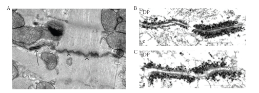
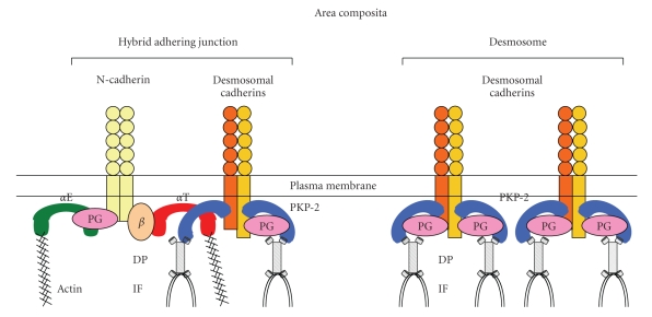
Adherens junctions and desmosomes are intercellular adhesive junctions and essential for the morphogenesis, differentiation, and maintenance of tissues that are subjected to high mechanical stress, including heart and skin. The different junction complexes are organized at the termini of the cardiomyocyte called the intercalated disc. Disruption of adhesive integrity via mutations in genes encoding desmosomal proteins causes an inherited heart disease, arrhythmogenic right ventricular cardiomyopathy (ARVC). Besides plakoglobin, which is shared by adherens junctions and desmosomes, other desmosomal components, desmoglein-2, desmocollin-2, plakophilin-2, and desmoplakin are also present in ultrastructurally defined fascia adherens junctions of heart muscle, but not other tissues. This mixed-type of junctional structure is termed hybrid adhering junction or area composita. Desmosomal plakophilin-2 directly interacts with adherens junction protein alphaT-catenin, providing a new molecular link between the cadherin-catenin complex and desmosome. The area composita only exists in the cardiac intercalated disc of mammalian species suggesting that it evolved to strengthen mechanical coupling in the heart of higher vertebrates. The cross-talk among different junctions and their implication in the pathogenesis of ARVC are discussed in this review.
Figures
References
-
- Basso C, Thiene G, Corrado D, Angelini A, Nava A, Valente M. Arrhythmogenic right ventricular cardiomyopathy: dysplasia, dystrophy, or myocarditis? Circulation. 1996;94(5):983–991. - PubMed
-
- Thiene G, Nava A, Corrado D, Rossi L, Pennelli N. Right ventricular cardiomyopathy and sudden death in young people. The New England Journal of Medicine. 1988;318(3):129–133. - PubMed
-
- Saffitz JE. Arrhythmogenic cardiomyopathy and abnormalities of cell-to-cell coupling. Heart Rhythm. 2009;6(8, supplement):S62–S65. - PubMed
Grants and funding
LinkOut - more resources
Full Text Sources
