. 2010 Jun 24;6(6):e1001003.
doi: 10.1371/journal.pgen.1001003.

Tandem E2F binding sites in the promoter of the p107 cell cycle regulator control p107 expression and its cellular functions

Affiliations

Tandem E2F binding sites in the promoter of the p107 cell cycle regulator control p107 expression and its cellular functions

Deborah L Burkhart et al. PLoS Genet. .

Abstract

The retinoblastoma tumor suppressor (Rb) is a potent and ubiquitously expressed cell cycle regulator, but patients with a germline Rb mutation develop a very specific tumor spectrum. This surprising observation raises the possibility that mechanisms that compensate for loss of Rb function are present or activated in many cell types. In particular, p107, a protein related to Rb, has been shown to functionally overlap for loss of Rb in several cellular contexts. To investigate the mechanisms underlying this functional redundancy between Rb and p107 in vivo, we used gene targeting in embryonic stem cells to engineer point mutations in two consensus E2F binding sites in the endogenous p107 promoter. Analysis of normal and mutant cells by gene expression and chromatin immunoprecipitation assays showed that members of the Rb and E2F families directly bound these two sites. Furthermore, we found that these two E2F sites controlled both the repression of p107 in quiescent cells and also its activation in cycling cells, as well as in Rb mutant cells. Cell cycle assays further indicated that activation of p107 transcription during S phase through the two E2F binding sites was critical for controlled cell cycle progression, uncovering a specific role for p107 to slow proliferation in mammalian cells. Direct transcriptional repression of p107 by Rb and E2F family members provides a molecular mechanism for a critical negative feedback loop during cell cycle progression and tumorigenesis. These experiments also suggest novel therapeutic strategies to increase the p107 levels in tumor cells.


Conflict of interest statement

The authors have declared that no competing interests exist.

Figures
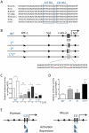
Figure 1
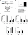
Figure 1. Regulation of the mouse p107 promoter through E2F binding sites in reporter assays.
(A) Conservation of the proximal p107 promoter across mammalian species. The two tandem consensus E2F binding sites (BS1 and BS2) are each indicated by a box. (B) Schematic representation of wild-type (WT), p107-1*, p107-2*, and p107-1*2* luciferase vectors. Transcription factor binding sites contained in this promoter region, as identified by sequence analysis, are indicated, as is the transcription start site (arrow). Black rectangular boxes indicate E2F consensus sites; white boxes indicate E2F consensus sites that are mutated. The inset represents the mutations (aaa) introduced in each site. (C) Relative luciferase activity expressed by the four constructs, co-transfected with CMV-E2F3 (+) or empty pCDNA (−), in cycling mESCs. For statistical analysis, each mutant construct was compared to the wild-type one and the effect of E2F3 on each construct was analyzed. (n = 3) (D) Relative luciferase activity in quiescent MEFs. (n = 15) (E) Comparison of the models for the regulation of the human and mouse p107 promoters by E2F based on reporter assays. Gradient triangles indicate the relative importance of each consensus E2F site to either activation or repression of p107.
Figure 2
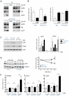
Figure 2. The E2F consensus binding sites in the p107 promoter are bound by E2F family members and control p107 expression in mESCs.
(A) Schematic representation of the targeting construct (top) used to knock-in mutations into the E2F sites in the endogenous p107 allele (bottom). Mutations in the E2F sites are indicated by asterisks. NeoR, neomycin resistance cassette; DTA, diphtheria toxin A. The black boxes indicate E2F consensus sites, the grey boxes indicate p107 exons. (B) Representative Southern analysis for a wild-type mESC clone (+/+) and a correctly targeted allele (+/*). Genomic DNA was digested by NdeI (N in Figure 1A) and a 5′ internal probe spanning the junction between the p107 intron and the NeoR cassette was used (black line in Figure 1A). (C) Schematic representation of the strategy used to generate homozygous mutant mESCs. (D) Sequencing analysis of wild-type and homozygous mutant mESCs. The knock-in mutant sequences are marked by boxes. (E) RT-qPCR analysis of p107 expression in wild-type and homozygous mutant cycling mESCs. p107 mRNA levels were calculated relative to TATA-binding protein (TBP). (n = 4) (F) Immunoblot analysis of p107 expression in mESCs. Tubulin expression is shown as a loading control. (G) Quantitative chromatin immunoprecipitation (ChIP) analysis of E2F3 (n = 5), E2F4 (n = 5), and p107 (n = 2) on the p107 promoter in wild-type (W) and homozygous mutant cycling mESCs. p16 antibodies serve as a negative control (n = 5). Fold enrichment was calculated over an unrelated DNA sequence (actin). The B-Myb promoter is shown as a control. The y-axis is plotted on a log2 scale.
Figure 3
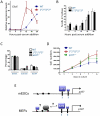
Figure 3. p107 repression in quiescent MEFs is mediated by the two E2F binding sites.
(A) mESCs targeted by the neomycin resistance cassette but retaining a wild-type p107 promoter and mESCs targeted by homozygous mutations into the distal (1*/1*) or both E2F sites (1*2*/1*2*) were injected to generate chimeric embryos. Wild-type, p107E2F-1*/1* and p107E2F-1*2*/1*2* MEFs derived from chimeric embryos were selected for Neomycin resistance to generate pure populations. (B) RT-qPCR analysis of p107 expression in quiescent wild-type and p107E2F-1*2*/1*2* MEFs. (n≥9) (C) Immunoblot analysis of p107 in the same conditions. Tubulin expression is shown as a loading control. (D) Quantitative ChIP analysis of E2F4, p107, and p130 binding on the p107 promoter in quiescent immortalized wild-type, p107E2F-1*/1*, and p107E2F-1*2*/1*2* MEFs. The B-Myb promoter is shown as a control. (n = 3) (E) Quantitative ChIP analysis of Rb binding to the p107 and Mcm3 promoters in cycling immortalized wild-type and p107E2F-1*2*/1*2* MEFs. Mouse IgG antibodies serve as a negative control. (n≥3) For (D,E), fold enrichment is calculated over actin and the y-axis is plotted on a log2 scale.
Figure 4
Figure 4. p107 levels increase in quiescent MEFs in the absence of Rb through transcriptional and post-translational mechanisms.
(A) Top: Representative immunoblot analysis of p107, Rb, and p130 expression after knockdown of Rb (shRb) or p130 (shp130-1 and shp130-2) as compared to empty vector in primary quiescent MEFs. Bottom: same experiment in p107E2F-1*2*/1*2* mutant MEFs. The asterisk shows a non-specific band that serves as a loading control; loading was also verified by Ponceau staining (not shown). (B) RT-qPCR analysis of Rb (left panel) and p107 (right panel) mRNA relative to TBP in quiescent wild-type and p107E2F-1*2*/1*2* MEFs infected with empty vector (−) or a vector to knock-down Rb (shRb). (n≥3) (C) Immunoblot analysis (left panel) of p107 levels in cells of the indicated genotypes infected with empty vector (−) or a vector to knock-down Rb (+). The E2F target PCNA serves as a positive control and β-Actin as a loading control. Protein quantification (right panel) is shown relative to β-Actin levels. p107 levels were not measured in p107 mutant cells (na). (n = 2) (D) Immunoblot analysis (left panel) of p107 levels in quiescent wild-type MEFs infected with empty vector (−) or a vector to knock-down Rb (shRb) and treated with cycloheximide (CHX) for 9 and 12 hours (hrs). Quantification (right panel) is shown relative to Tubulin levels. (n = 2) (E) RT-qPCR analysis of B-Myb (left panel), E2F1 (center panel), and Cdc6 (right panel) mRNA levels relative to TBP in quiescent primary wild-type, p107E2F-1*2*/1*2*, and p107−/− MEFs after knockdown of Rb as in B. For statistical analysis, each MEF genotype was compared to the wild-type one by an unpaired Student's t-test, and the effect of Rb knockdown on each genotype was analyzed by a paired Student's t-test. (n≥3)
Figure 5
Figure 5. E2F binding sites mediate activation of the p107 promoter in cycling cells.
(A) RT-qPCR analysis of p107 mRNA relative to TBP in asynchronously cycling primary wild-type and p107E2F-1*2*/1*2* MEFs. (n = 12) (B) Immunoblot analysis (left panel) of p107 expression in wild-type and p107E2F-1*2*/1*2* MEFs as in A. Tubulin expression is shown as a loading control. p107 protein quantification (right panel) is shown relative to Tubulin levels. (n = 3) (C) Representative example of Hoescht33342 staining of asynchronously cycling MEFs showing G1 and S phase populations; wild-type and mutant cells have similar profiles (data not shown). (D) RT-qPCR analysis of immortalized WT, p107E2F-1*/1* and p107E2F-1*2*/1*2* MEFs. For each genotype, G0 samples were collected after at least three days of serum starvation. Asynchronous cells were stained with Hoechst33342 and sorted by their DNA content into G1 and S-phase samples. (n≥2) (E) and (F) RT-qPCR analysis of primary wild-type and p107E2F-1*2*/1*2* MEFs that have been synchronized in G0 by serum starvation. DMEM supplemented with 20% serum was added at time 0, and extracts were collected at 10 hrs, 16 hrs, 22 hrs, and 28 hrs post-stimulation. (E) p107 mRNA and (F) Cdc6 mRNA. n≥8 for both genotypes at all time points. (G) Percentage of cells in S-phase in primary MEFs collected during cell-cycle re-entry as in E. and F. Percentages were calculated by BrdU/PI analysis (n = 3). (H) Immunoblot analysis of p107 protein expression in primary wild-type and p107E2F-1*2*/1*2* MEF extracts collected at 0 hr, 8 hrs, 12 hrs, 16 hrs, 20 hrs, and 24 hrs post-stimulation with 20% serum. MCM6 expression is shown as a positive control for cell cycle re-entry, and Tubulin levels are shown as a loading control. Note that the second, slowly migrating form of p107 at later time points probably reflects p107 phosphorylation during S phase.
Figure 6
Figure 6. Altered p107 expression affects cellular proliferation.
(A,B) Immortalized wild-type and p107E2F-1*2*/1*2* MEFs were synchronized in G0 through at least three days of serum starvation. DMEM supplemented with 20% BGS was used to stimulate cell-cycle entry. Extracts were collected at the number of hours indicated post-serum stimulation. (A) RT-qPCR analysis of Cdc6 mRNA in wild-type and p107E2F-1*2*/1*2* MEFs. (n = 3) (B) Percentage of cells in S-phase, as determined by BrdU/PI staining, at the indicated time points. (n≥4) (C) Cell-cycle profiles of asynchronous primary wild-type, p107E2F-1*2*/1*2*, and p107−/− MEFs. Percentages of cells in each phase were determined by BrdU/PI staining. (n≥2) (D) Cellular proliferation of primary wild-type, p107E2F-1*2*/1*2*, and p107−/− MEFs. Equal numbers of cells were plated at day 0. Cells were then counted every other day from day 1 to day 9 post-plating. For statistical analysis, p107E2F-1*2*/1*2* cells were compared to wild-type cells at each time point. (n≥13) (E) Model for the context-dependent regulation of p107 transcription by E2F family members. In cycling mESCs, activating members of the E2F family such as E2F3 bind to the p107 promoter mostly through the distal consensus E2F binding site (site 1). In quiescent MEFs, binding of the E2F4 repressor is also largely dependent on the presence of the distal consensus site. However, E2F4 may also be recruited to the p107 promoter through interactions with other transcription factors and/or by binding to other DNA sequences. The size of the E2F boxes indicates the relative binding activity.

Similar articles

Cited by

References

    1. Burkhart DL, Sage J. Cellular mechanisms of tumour suppression by the retinoblastoma gene. Nat Rev Cancer. 2008;8:671–682. - PMC - PubMed
    1. Classon M, Dyson N. p107 and p130: versatile proteins with interesting pockets. Exp Cell Res. 2001;264:135–147. - PubMed
    1. Trimarchi JM, Lees JA. Sibling rivalry in the E2F family. Nat Rev Mol Cell Biol. 2002;3:11–20. - PubMed
    1. Ewen ME, Xing YG, Lawrence JB, Livingston DM. Molecular cloning, chromosomal mapping, and expression of the cDNA for p107, a retinoblastoma gene product-related protein. Cell. 1991;66:1155–1164. - PubMed
    1. Yu CL, Tucker MA, Abramson DH, Furukawa K, Seddon JM, et al. Cause-specific mortality in long-term survivors of retinoblastoma. J Natl Cancer Inst. 2009;101:581–591. - PMC - PubMed

Publication types

Substances