. 2010 Jun 23;5(6):e11195.
doi: 10.1371/journal.pone.0011195.

Identification of conserved regions and residues within Hedgehog acyltransferase critical for palmitoylation of Sonic Hedgehog

Affiliations

Identification of conserved regions and residues within Hedgehog acyltransferase critical for palmitoylation of Sonic Hedgehog

John A Buglino et al. PLoS One. .

Abstract

Background: Sonic hedgehog (Shh) is a palmitoylated protein that plays key roles in mammalian development and human cancers. Palmitoylation of Shh is required for effective long and short range Shh-mediated signaling. Attachment of palmitate to Shh is catalyzed by Hedgehog acyltransferase (Hhat), a member of the membrane bound O-acyl transferase (MBOAT) family of multipass membrane proteins. The extremely hydrophobic composition of MBOAT proteins has limited their biochemical characterization. Except for mutagenesis of two conserved residues, there has been no structure-function analysis of Hhat, and the regions of the protein required for Shh palmitoylation are unknown.

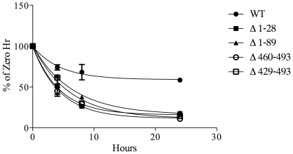
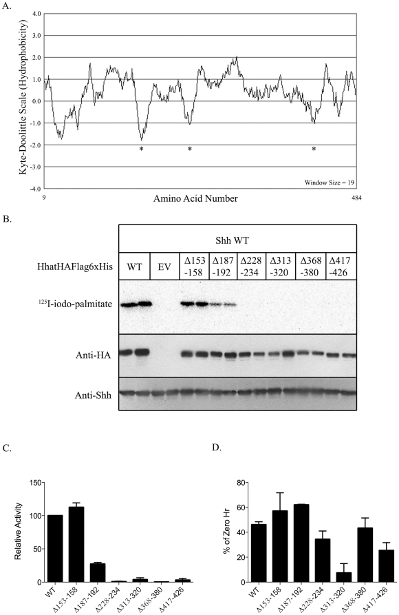
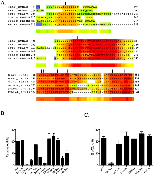
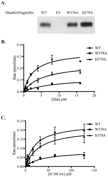
Methodology/principal findings: Here we undertake a systematic approach to identify residues within Hhat that are required for protein stability and/or enzymatic activity. We also identify a second, novel MBOAT homology region (residues 196-234) that is required for Hhat activity. In total, ten deletion mutants and eleven point mutants were generated and analyzed. Truncations at the N- and C-termini of Hhat yielded inactive proteins with reduced stability. Four Hhat mutants with deletions within predicted loop regions and five point mutants retained stability but lost palmitoylation activity. We purified two point mutants, W378A and H379A, with defective Hhat activity. Kinetic analyses revealed alterations in apparent K(m) and V(max) for Shh and/or palmitoyl CoA, changes that likely explain the catalytic defects observed for these mutants.

Conclusions/significance: This study has pinpointed specific regions and multiple residues that regulate Hhat stability and catalysis. Our findings should be applicable to other MBOAT proteins that mediate lipid modification of Wnt proteins and ghrelin, and should serve as a model for understanding how secreted morphogens are modified by palmitoyl acyltransferases.


Conflict of interest statement

Competing Interests: The authors have declared that no competing interests exist.

Figures

Figure 1
Figure 1. N and C Terminal truncation mutants of Hhat lack PAT activity.
A. Transmembrane topology model of Hhat generated using the TMHMM v. 2.0 Server. Gray bars indicate predicted transmembrane helices. Arrows denote approximate truncation points. B. COS-1 cells were transfected with the indicated constructs and labeled with 125I-iodo-palmitate for 4h. Cell lysates were analyzed directly by Western blotting or after immunoprecipitation of Shh. Upper panel: 125I-iodo-palmitate incorporation into immunoprecipitated Shh as detected by phosphorimaging. Lower panels: Western blots of the same extracts probed with anti-HA, anti-Shh, and anti-β-tubulin (β-Tub) antibodies. EV, empty vector. C. An in vitro palmitoylation assay was performed with P100 membranes generated from 293FT cells transfected with WT and mutant Hhat constructs. Upper panel: 125I-iodo-palmitate incorporation into Shh detected by phosphorimaging. Lower panels: Western blots of the same samples probed with anti-HA and anti-Shh antibodies. D. Quantification of the experiments in panels B and C. Levels of 125I-iodo-palmitate incorporation were corrected for Hhat protein expression and normalized to WT Hhat levels (100%). Each bar represents the average of three experiments and is expressed as the percent of WT activity (set to 100%).
Figure 2
Figure 2. Stability of Hhat truncation mutants.
COS-1 cells transfected with the indicated Hhat constructs were incubated in DMEM supplemented with 10% FBS, 100 µg/ml cycloheximide, and 40 µg/ml chloramphenicol. At each indicated time point, cells were lysed and subjected to SDS-PAGE and Western blotting with anti-HA antibodies. The amount of HA signal at each time point was determined using ImageJ software. Data are expressed as percent of 0 h controls, which were set to 100%. Experiments were carried out in duplicate and repeated three times. Values for the percentage of Hhat protein remaining at 24 h were: WT, 58%; Δ1–28, 17%, Δ1–89 19%, Δ460–493 11%, Δ429–493 14%. Estimated half-lives for the mutants were 3–7 h, compared to >20 h for WT Hhat.
Figure 3
Figure 3. PAT activity and stability measurements of Hhat constructs containing deletions within predicted loop regions.
A. Kyte-Doolittle hydropathy plot of Hhat with the window size set at 19. Asterisks indicate regions of high hydrophilicity that were targeted for mutagenesis. B. Representative in vitro palmitoylation assay. P100 membranes isolated from 293FT cells expressing the indicated Hhat constructs were reacted with Shh and 125I-iodo-palmitate CoA as described in Materials and Methods. EV, empty vector. C. Quantification of the palmitoylation assays performed three times. Levels of 125I-iodo-palmitate incorporation were corrected for Hhat protein expression and normalized to WT Hhat (100%). Data are expressed as a percent of WT activity. D. Relative stability of Hhat deletion mutants after 24 h incubation with cycloheximide and chloramphenicol, as described in Figure 2. Data are expressed as a percentage of 0 h controls. Experiments were performed in duplicate and repeated three times.
Figure 4
Figure 4. Identification of MBOAT Homology Regions.
A. Multiple sequence alignment of MBOAT family members that acylate protein substrates generated using the TCoffee server. The sequence from GUP-1/Hhat-like protein was included based on its high homology to Hhat and the functional characterization of GUP-1 from the yeast S. cerevisiae . Arrows indicate residues mutated to alanine. B. Quantification of in vitro palmitoylation assays performed three times. Levels of 125I-iodo-palmitate incorporation were corrected for Hhat protein expression and normalized to WT Hhat (100%). Data is expressed as a percent of WT activity. C. Relative stability of Hhat point mutants after 24 h incubation with cycloheximide and chloramphenicol. Data are expressed as percent of 0 h controls. Experiments were carried out in duplicate and repeated three times.
Figure 5
Figure 5. Kinetic analyses of purified Hhat mutants.
A. The indicated Hhat constructs were expressed in and purified from 293FT cells as described in Materials and Methods. Aliquots of each purified protein were analyzed by SDS-PAGE and Western blotting using anti-Flag antibody. EV, empty vector. B. Each purified HhatHAFlagHis construct was incubated with Shh at the indicated concentration in the presence of 100 µM 125I-iodo-palmitoyl CoA for 1 h at room temperature. C. Each purified HhatHAFlagHis construct was incubated with 125I-iodo-palmitoyl CoA at the indicated concentration in the presence of 40 µM Shh. Shh protein bands were excised from dried gels and the amount of 125I-iodo-palmitate incorporation was determined by gamma counting. Graphs represent the average of three experiments corrected for non-specific incorporation of 125I-iodo-palmitate as described previously .

Similar articles

Cited by

References

    1. Ho KS, Scott MP. Sonic hedgehog in the nervous system: functions, modifications and mechanisms. Curr Opin Neurobiol. 2002;12:57–63. - PubMed
    1. Fuccillo M, Joyner AL, Fishell G. Morphogen to mitogen: the multiple roles of hedgehog signalling in vertebrate neural development. Nat Rev Neurosci. 2006;7:772–783. - PubMed
    1. McMahon AP, Ingham PW, Tabin CJ. Developmental roles and clinical significance of hedgehog signaling. Curr Top Dev Biol. 2003;53:1–114. - PubMed
    1. Lee JD, Kraus P, Gaiano N, Nery S, Kohtz J, et al. An acylatable residue of Hedgehog is differentially required in Drosophila and mouse limb development. Dev Biol. 2001;233:122–136. - PubMed
    1. Pasca di Magliano M, Hebrok M. Hedgehog signalling in cancer formation and maintenance. Nat Rev Cancer. 2003;3:903–911. - PubMed

Publication types

LinkOut - more resources