

Shed blood-derived cells from total hip arthroplasty have osteoinductive potential: a pilot study
- PMID: 20585911
- PMCID: PMC3049633
- DOI: 10.1007/s11999-010-1444-z
Shed blood-derived cells from total hip arthroplasty have osteoinductive potential: a pilot study
Abstract
Background: Cell therapy using autologous cells has been used in the treatment of various medical conditions. The mononuclear cell (MNC) fraction of bone marrow (BM) contains stem/progenitor cells that could contribute to osteogenesis and angiogenesis.
Questions/purposes: We asked whether MNCs derived from intraoperative shed blood (SB), consisting of peripheral blood and BM, have osteoinductive and angiogenic potential.
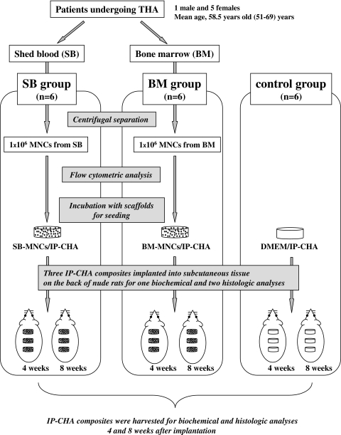
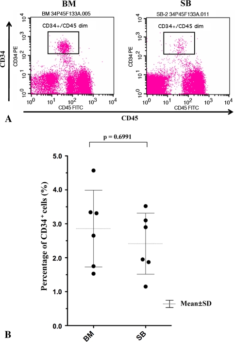
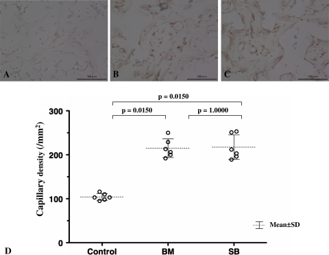
Methods: We harvested SB and BM from six patients undergoing THA. Isolated MNCs from SB and BM were analyzed by flow cytometry to evaluate the CD34(+) cell fraction and 1 × 10(6) cells were seeded on an interconnective porous calcium hydroxyapatite ceramic (IP-CHA) and transplanted in the backs of athymic rats. IP-CHAs without cells were transplanted as controls and all composites were harvested after 4 and 8 weeks. Osteoinductive potential was evaluated by histologic observation, immunohistochemistry, and enzyme-linked immunosorbent assay (ELISA) using anti-osteocalcin (OC) antibodies qualitatively and quantitatively. To evaluate angiogenic potential, capillary density was measured by immunohistochemistry using Isolectin B4 4 weeks after implantation.
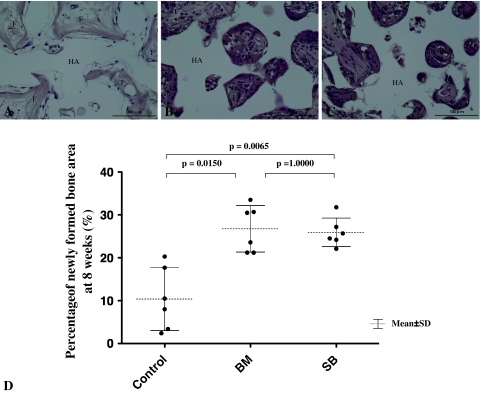
Results: We found that CD34(+) cells existed in SB-MNCs and there was a trend toward lower frequency compared with BM-MNCs. Histologic osteoinduction, OC expression, and capillary density were increased by transplantation of MNCs from SB. Similar results were achieved with MNCs from BM.
Conclusions: MNCs from SB have equivalent osteoinductive and angiogenic potential compared with those from BM.
Clinical relevance: SB could be an attractive source for isolation of MNCs, enhancing osteoinduction and neovascularization, to augment the reconstruction of skeletal defects.
Figures


Similar articles
-
Therapeutic angiogenesis of bone marrow mononuclear cells (MNCs) and peripheral blood MNCs: transplantation for ischemic hindlimb.Ann Vasc Surg. 2008 Mar;22(2):238-47. doi: 10.1016/j.avsg.2007.07.037. Epub 2008 Feb 20. Ann Vasc Surg. 2008. PMID: 18083329
-
Improvement of collateral perfusion and regional function by implantation of peripheral blood mononuclear cells into ischemic hibernating myocardium.Arterioscler Thromb Vasc Biol. 2002 Nov 1;22(11):1804-10. doi: 10.1161/01.atv.0000039168.95670.b9. Arterioscler Thromb Vasc Biol. 2002. PMID: 12426208
-
Age-related BM-MNC dysfunction hampers neovascularization.Mech Ageing Dev. 2007 Sep;128(9):511-6. doi: 10.1016/j.mad.2007.06.009. Epub 2007 Jul 4. Mech Ageing Dev. 2007. PMID: 17688912
-
Clinical efficacy of autologous stem cell transplantation for the treatment of patients with type 2 diabetes mellitus: a meta-analysis.Cytotherapy. 2015 Jul;17(7):956-68. doi: 10.1016/j.jcyt.2015.02.014. Epub 2015 Mar 29. Cytotherapy. 2015. PMID: 25824289 Review.
-
Transplantation of endothelial progenitor cells for therapeutic neovascularization.Cardiovasc Radiat Med. 2002 Jul-Dec;3(3-4):221-5. doi: 10.1016/s1522-1865(03)00082-9. Cardiovasc Radiat Med. 2002. PMID: 12974378 Review.
References
-
- Bartsch T, Brehm M, Zeus T, Strauer BE. Autologous mononuclear stem cell transplantation in patients with peripheral occlusive arterial disease. J Cardiovasc Nurs. 2006;21:430–432. - PubMed
-
- Bragdon CR, Doherty AM, Rubash HE, Jasty M, Li XJ, Seeherman H, Harris WH. The efficacy of BMP-2 to induce bone ingrowth in a total hip replacement model. Clin Orthop Relat Res. 2003;417:50–61. - PubMed
MeSH terms
Substances
LinkOut - more resources
Full Text Sources
Medical
