. 2010 Oct;13(5):581-8.
doi: 10.1089/rej.2009.1011. Epub 2010 Jun 29.

NT-020, a natural therapeutic approach to optimize spatial memory performance and increase neural progenitor cell proliferation and decrease inflammation in the aged rat

Affiliations

NT-020, a natural therapeutic approach to optimize spatial memory performance and increase neural progenitor cell proliferation and decrease inflammation in the aged rat

S Acosta et al. Rejuvenation Res. 2010 Oct.

Abstract

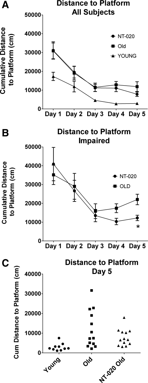
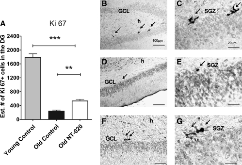
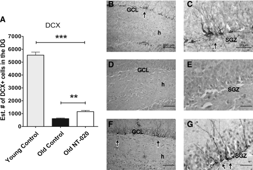
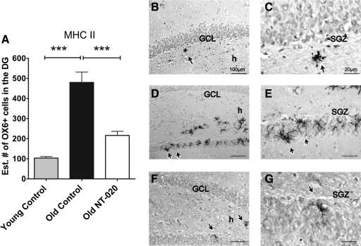
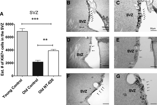
The process of aging is linked to oxidative stress, microglial activation, and proinflammatory factors, which are known to decrease cell proliferation and limit neuroplasticity. These factors may lead the transition from normal aging to more severe cognitive dysfunction associated with neurodegenerative diseases. We have shown that natural compounds such as polyphenols from blueberry and green tea and amino acids like carnosine are high in antioxidant and antiinflammatory activity that decreases the damaging effects of reactive oxygen species (ROS), in the blood, brain, and other tissues of the body. Furthermore, we have shown that the combination of these nutrients (called NT-020) creates a synergistic effect that promotes the proliferation of stem cells in vitro and in vivo. In the current study, we examined the effects of NT-020 on neurogenesis and performance on a Morris water maze (MWM). Aged (20-month-old) male Fischer 344 rats were treated with 135.0 mg/kg per day (n = 13) of NT-020. Young (3-month-old) (n = 10) and aged (20-month-old) (n = 13) control male Fischer 344 rats were treated with water by oral gavage. All groups were treated for a period of 4 weeks. Although there was no difference in performance in the MWM when comparing all aged rats, when the data for aged impaired rats were compared, there was a significant difference between groups on the last day of training with the treatment group performing better than controls. Using the cell cycle-regulating protein (Ki67), doublecortin (DCX), and OX6 antibody markers, cell proliferation, neurogenesis, and microglial activation were estimated in the dentate gyrus (DG) of young and aged animals. Cell proliferation was also examined in the subventricular zone (SVZ). A decreased number of OX6 MHC II-positive cells, increased neurogenesis, and increased number of proliferating cells were found in rats treated with NT-020 in comparison with aged control rats. In sum, NT-020 may promote health, proliferation, and maintenance of neurons in the age animals and exert antiinflammatory actions that promote function in the aged stem cell niche.


Figures

FIG. 1.
FIG. 1.
Cumulative distance to platform. (A) Morris Water maze learning acquisition performance of all subjects. When learning over days is graphed for all subjects, there is a clear age difference where young rats outperform the aged rats; then by days 4 and 5, a difference begins to emerge between the treatment group and the control aged group. (B) When the highest scoring 40% of rats are examined as the aged impaired (AI) groups for both the control and NT-020 treatment, there is a significant difference between groups on days 5 (one-way analysis of variance [ANOVA] followed by Bonferroni analysis; overall F = 16.55, degrees of freedom [df ] = 2, 12 post hoc for aged control vs. treatment p < 0.05; young vs. NT-020 is not significantly different) and distance to platform (ANOVA, F = 22.6 df = 2, 12, aged control vs. aged NT-020 p < 0.01). (C) Individual rat performance on day 5 of training is shown to demonstrate the wide variance of data within subjects and that there was lower variance in the NT-020-treated group.
FIG. 2.
FIG. 2.
(A) Ki 67+ cells (cell cycle marker) present in the subgranular zone (SGZ) of the dentate gyrus (DG) of young (4-month-old, n = 10) and aged (21-month-old, n = 13) rats quantified using stereology methods. Asterisks denote a significant age-related decrease in cell proliferation, demonstrating a significant increase of cell proliferation in the NT-020 oral gavage treatment group compared with age-matched controls. There is an overall age-related decrease in Ki67+ cells with age. (One-way analysis of variance [ANOVA] F = 157.6, degrees of freedom [df ] = 2, 32; followed by Bonferroni post hoc values [***] p = 0.005, [**] p < < 0.05). Photomicrographs depicting representative sections of Ki67 staining in the granular cell layer (GCL) and SGZ of the DG of the hippocampus (arrows) in young rats (B,C), in control aged rats (D,E), and in NT-020 aged, treated rats (F,G). Scale bars for B, D, F, 100 μm; C, E, G, 20 μm. h indicates the hilus of the dentate gyrus.
FIG. 3.
FIG. 3.
Doublecortin+ cells (DCX) present in granule cell layer of the dentate gyrus (DG) of young (4-month-old, n = 10) and aged rats (21-month-old, n = 13). Quantification of DCX shows a significant increase of DCX+ cells in the DG of aged rats treated with NT-020 for 1 month in comparison with aged rats treated with water oral gavage (one-way analysis of variance [ANOVA] F = 487.9, degrees of freedom [df ] = 2, 24; Bonferroni post hoc values [***] p < 0.005, [**] p < 0.05). Photomicrographs depicting representative sections of DCX staining in the granular cell layer (GCL) and subgranular zone (SGZ) of the DG of the hippocampus (arrows) in young rats (B,C), in control aged rats (D,E), and NT-020 aged, treated rats (F,G). Scale bars for B, D, F, 100 μm; C, E, G, 20 μm. h indicates the hilus of the dentate gyrus.
FIG. 4.
FIG. 4.
(A) OX6+ cells (MHC class II) present in the granule cell layer of the dentate gyrus (DG) (arrows) of young (4-month-old, n = 10) and aged (21-month-old, n = 13) rats counted using stereology. Asterisks denote a significant age-related increase in MHC class II–expressing cells, demonstrating a significant decrease in MHC class II expression in the NT-020 oral gavage treatment group compared with age-matched controls. There is an overall age-related increase in OX6 expression in cells with age. (One-way analysis of variance [ANOVA] F = 31.63, degrees of freedom [df ] = 2, 32; followed by Bonferonni post hoc values [***] p < 0.005, [***] p < 0.05). Photomicrographs depicting representative sections of OX6 staining in the granular cell layer (GCL) and subgranular zone (SGZ) of the DG of the hippocampus (arrows) in young rats (B,C), in control aged rats (D,E), and NT-020 aged, treated rats (F,G). Scale bars, B, D, F, 100 μm; C, E, G, I 20 μm. h indicates the hilus of the dentate gyrus.
FIG. 5.
FIG. 5.
(A) Ki67+ cells (cell cycle marker) present in the subventricular zone (SVZ) of young (4-month-old, n = 10) and aged (21-month-old, n = 13) rats counted using stereology. Asterisks denote a significant age-related decline in Ki67-expressing cells, demonstrating a significant increase in number of cells expressing Ki67 staining in the NT-020 treatment group compared with age-matched controls. There is an overall age-related decrease in Ki67+ cells in the SVZ with age. (One-way analysis of variance [ANOVA] F = 52.82, degrees of freedom [df ] = 2, 24; followed by Bonferroni post hoc values [***] p < 0.005, [**] p < 0.05). Photomicrographs depicting representative sections of OX6 staining in the granular cell layer (GCL) and subgranular zone (SGZ) of the dentate gyrus (DG) of the hippocampus (arrows) in young rats (B,C), in control aged rats, (D,E), and NT-020 aged, treated rats (F,G). Scale bars, B, D, F, 100 μm; scale bar, C, E, G, 50 μm. h indicates the hilus of the dentate gyrus.

References

    1. Floyd RA. Hensley K. Oxidative stress in brain aging. Implications for therapeutics of neurodegenerative diseases. Neurobiol Aging. 2002;23:795–807. - PubMed
    1. Arnaiz E. Almkvist O. Neuropsychological features of mild cognitive impairment and preclinical Alzheimer's disease. Acta Neurol Scand Suppl. 2003;179:34–41. - PubMed
    1. Bickford PC. Gould T. Briederick L. Chadman K. Pollock A. Young D. Shukitt-Hale B. Joseph J. Antioxidant-rich diets improve cerebellar physiology and motor learning in aged rats. Brain Res. 2000;866:211–217. - PubMed
    1. Gemma C. Mesches MH. Sepesi B. Choo K. Holmes DB. Bickford PC. Diets enriched in foods with high antioxidant activity reverse age-induced decreases in cerebellar beta-adrenergic function and increases in proinflammatory cytokines. J Neurosci. 2002;22:6114–120. - PMC - PubMed
    1. Liu J. Atamna H. Kuratsune H. Ames BN. Delaying brain mitochondrial decay and aging with mitochondrial antioxidants and metabolites. Ann NY Acad Sci. 2002;959:133–166. - PubMed

Publication types

MeSH terms