

Pituitary hCG production and cerebral tuberculosis mimicking disease progression during chemotherapy for an advanced ovarian germ cell tumour
- PMID: 20587067
- PMCID: PMC2909206
- DOI: 10.1186/1471-2407-10-338
Pituitary hCG production and cerebral tuberculosis mimicking disease progression during chemotherapy for an advanced ovarian germ cell tumour
Abstract
Background: Ovarian germ cell tumours (OGCT) are rare but are usually curable with chemotherapy, even when presenting with advanced disease. The majority of OGCT produce the tumour markers, hCG and/or AFP which can be helpful in the diagnosis and monitoring the response to treatment.
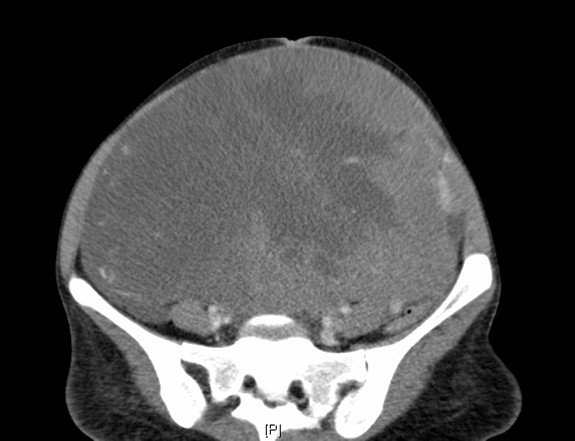
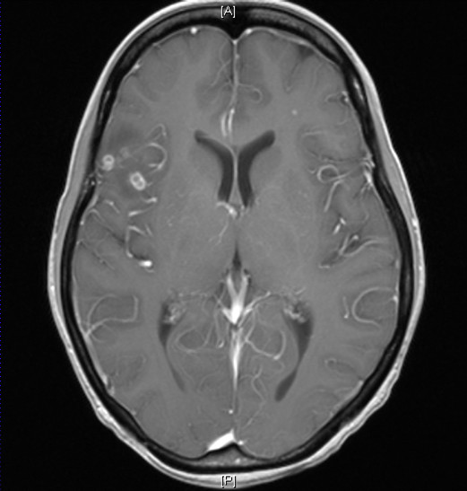
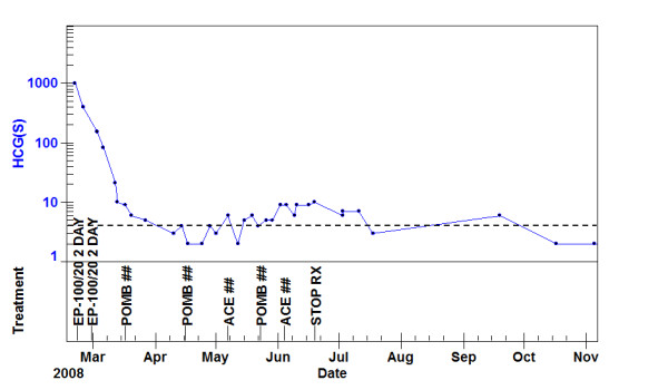
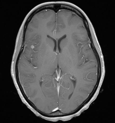
Case presentation: In this case of a 36 year old woman, the elevated hCG level at presentation was helpful in making a clinical diagnosis of OGCT in a patient too unwell to permit a tissue diagnosis. Cisplatin based combination chemotherapy produced an initial normalisation of the hCG level, but later in treatment the patient developed new cerebral lesions and a rising serum hCG suggestive of disease progression. Further investigations suggested that the CNS lesions were cerebral TB and that the low levels of hCG elevations was likely to be pituitary in origin. Chemotherapy treatment was continued along with anti-tuberculous therapy and 24 months after successful completion of therapy the patient remains disease free.
Conclusions: In the treatment of cancer patients it may be helpful to consider the potential non-malignant causes of new CNS lesions and that low hCG elevations may result from physiology rather than pathology in selected cases.
Figures
Similar articles
-
Ovarian germ cell tumours: a 17-year study in a single unit.Eur J Pediatr Surg. 2009 Apr;19(2):96-100. doi: 10.1055/s-0029-1202372. Epub 2009 Apr 9. Eur J Pediatr Surg. 2009. PMID: 19360543
-
The UK Children's Cancer Study Group: testicular malignant germ cell tumours 1979-1988.J Pediatr Surg. 1990 Apr;25(4):406-10. doi: 10.1016/0022-3468(90)90381-i. J Pediatr Surg. 1990. PMID: 1691781
-
Chemotherapy of germ-cell ovarian tumours: first-line treatment with etoposide, bleomycin and cisplatin or carboplatin.Eur J Cancer Clin Oncol. 1987 May;23(5):469-74. doi: 10.1016/0277-5379(87)90305-1. Eur J Cancer Clin Oncol. 1987. PMID: 2443358
-
False-positive serum human chorionic gonadotropin (HCG) in a male patient with a malignant germ cell tumor of the testis: a case report and review of the literature.Oncologist. 2008 Nov;13(11):1149-54. doi: 10.1634/theoncologist.2008-0159. Epub 2008 Nov 4. Oncologist. 2008. PMID: 18984875 Review.
-
Paraneoplastic Hyperthyroidism Secondary to a Chemotherapy-Induced Surge in β-hCG in a Patient with Non-Seminomatous Germ Cell Tumor.Hawaii J Health Soc Welf. 2024 Feb;83(2):45-47. Hawaii J Health Soc Welf. 2024. PMID: 38344694 Free PMC article. Review.
Cited by
-
Pituitary as a Source of HCG: Residual Levels After Bilateral Testicular Tumor Removal.J Investig Med High Impact Case Rep. 2019 Jan-Dec;7:2324709619841414. doi: 10.1177/2324709619841414. J Investig Med High Impact Case Rep. 2019. PMID: 31010310 Free PMC article. Review.
References
-
- Murugaesu N, Schmid P, Dancey G, Agarwal R, Holden L, McNeish I, Savage PM, Newlands ES, Rustin GJ, Seckl MJ. Malignant ovarian germ cell tumors: identification of novel prognostic markers and long-term outcome after multimodality treatment. J Clin Oncol. 2006;24(30):4862–6. doi: 10.1200/JCO.2006.06.2489. - DOI - PubMed
-
- Fosså SD, Stenning SP, Gerl A, Horwich A, Clark PI, Wilkinson PM, Jones WG, Williams MV, Oliver RT, Newlands ES, Mead GM, Cullen MH, Kaye SB, Rustin GJ, Cook PA. Prognostic factors in patients progressing after cisplatin-based chemotherapy for malignant non-seminomatous germ cell tumours. Br J Cancer. 1999;80(9):1392–9. doi: 10.1038/sj.bjc.6690534. - DOI - PMC - PubMed
Publication types
MeSH terms
Substances
LinkOut - more resources
Full Text Sources
Medical
