

Nuclear localization of orphan receptor protein kinase (Ror1) is mediated through the juxtamembrane domain
- PMID: 20587074
- PMCID: PMC2907318
- DOI: 10.1186/1471-2121-11-48
Nuclear localization of orphan receptor protein kinase (Ror1) is mediated through the juxtamembrane domain
Abstract
Background: Several receptor tyrosine kinases (RTKs) such as EGFR, FGFR, TRK, and VEGFR are capable of localizing in the cell nucleus in addition to their usual plasma membrane localization. Recent reports also demonstrate that nuclear-localized RTKs have important cellular functions such as transcriptional activation. On the basis of preliminary bioinformatic analysis, additional RTKs, including receptor tyrosine kinase-like orphan receptor 1 (Ror1) were predicted to have the potential for nuclear subcellular localization. Ror1 is a receptor protein tyrosine kinase that modulates neurite growth in the central nervous system. Because the nuclear localization capability of the Ror1 cytoplasmic domain has not been reported, we examined the cellular expression distribution of this region.
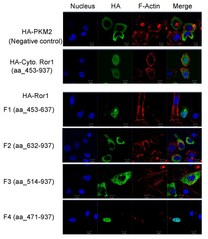
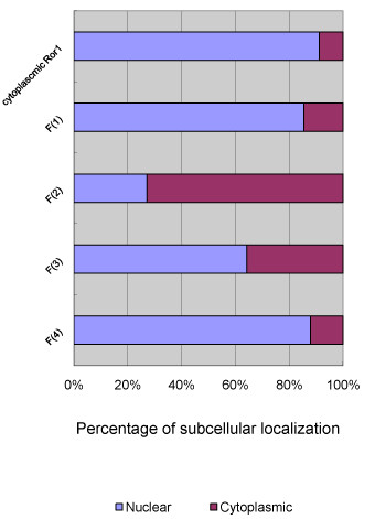
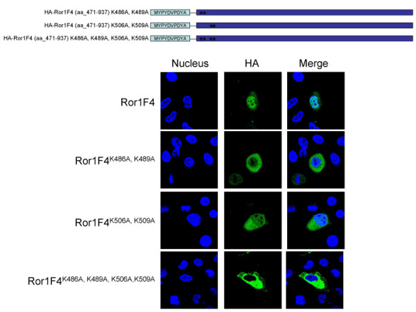
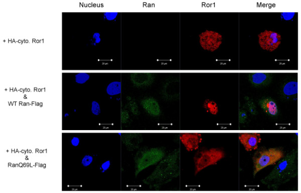
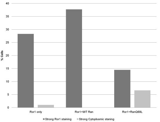
Results: The Ror1 cytoplasmic region was amplified and cloned into reporter constructs with fluorescent tags. Following transfection, the nuclear distribution patterns of transiently expressed fusion proteins were observed. Serial deletion constructs were then used to map the juxtamembrane domain of Ror1 (aa_471-513) for this nuclear translocation activity. Further site-directed mutagenesis suggested that a KxxK-16 aa-KxxK sequence at residues 486-509 is responsible for the nuclear translocation interaction. Subsequent immunofluorescence analysis by cotransfection of Ran and Ror1 implied that the nuclear translocation event of Ror1 might be mediated through the Ran pathway.
Conclusions: We have predicted several RTKs that contain the nuclear localization signals. This is the first report to suggest that the juxtamembrane domain of the Ror1 cytoplasmic region mediates the translocation event. Ran GTPase is also implicated in this event. Our study might be beneficial in future research to understand the Ror1 biological signaling pathway.
Figures

Similar articles
-
Receptor tyrosine kinase-like orphan receptor 1, a target of NKX2-1/TTF-1 lineage-survival oncogene, inhibits apoptosis signal-regulating kinase 1-mediated pro-apoptotic signaling in lung adenocarcinoma.Cancer Sci. 2016 Feb;107(2):155-61. doi: 10.1111/cas.12858. Epub 2016 Feb 8. Cancer Sci. 2016. PMID: 26661061 Free PMC article.
-
ROR1-CAVIN3 interaction required for caveolae-dependent endocytosis and pro-survival signaling in lung adenocarcinoma.Oncogene. 2019 Jun;38(26):5142-5157. doi: 10.1038/s41388-019-0785-7. Epub 2019 Mar 20. Oncogene. 2019. PMID: 30894682
-
Stat3 activates the receptor tyrosine kinase like orphan receptor-1 gene in chronic lymphocytic leukemia cells.PLoS One. 2010 Jul 29;5(7):e11859. doi: 10.1371/journal.pone.0011859. PLoS One. 2010. PMID: 20686606 Free PMC article.
-
Receptor tyrosine kinase-like orphan receptor 1: a novel target for cancer immunotherapy.Expert Opin Ther Targets. 2015 Jul;19(7):941-55. doi: 10.1517/14728222.2015.1025753. Epub 2015 Apr 2. Expert Opin Ther Targets. 2015. PMID: 25835638 Review.
-
The receptor tyrosine kinase ROR1--an oncofetal antigen for targeted cancer therapy.Semin Cancer Biol. 2014 Dec;29:21-31. doi: 10.1016/j.semcancer.2014.07.005. Epub 2014 Jul 25. Semin Cancer Biol. 2014. PMID: 25068995 Review.
Cited by
-
Proteolytic cleavage, trafficking, and functions of nuclear receptor tyrosine kinases.FEBS J. 2015 Oct;282(19):3693-721. doi: 10.1111/febs.13342. Epub 2015 Jul 4. FEBS J. 2015. PMID: 26096795 Free PMC article. Review.
-
The NAE Pathway: Autobahn to the Nucleus for Cell Surface Receptors.Cells. 2019 Aug 16;8(8):915. doi: 10.3390/cells8080915. Cells. 2019. PMID: 31426451 Free PMC article. Review.
-
Wnt/β-catenin signaling during early vertebrate neural development.Dev Neurobiol. 2017 Nov;77(11):1239-1259. doi: 10.1002/dneu.22517. Epub 2017 Aug 21. Dev Neurobiol. 2017. PMID: 28799266 Free PMC article. Review.
-
Targeting the Receptor Tyrosine Kinase ROR1 by Small Molecules.Handb Exp Pharmacol. 2021;269:75-99. doi: 10.1007/164_2021_535. Handb Exp Pharmacol. 2021. PMID: 34490515
-
The role of Ryk and Ror receptor tyrosine kinases in Wnt signal transduction.Cold Spring Harb Perspect Biol. 2014 Feb 1;6(2):a009175. doi: 10.1101/cshperspect.a009175. Cold Spring Harb Perspect Biol. 2014. PMID: 24370848 Free PMC article. Review.
References
-
- Hung LY, Tseng JT, Lee YC, Xia W, Wang YN, Wu ML, Chuang YH, Lai CH, Chang WC. Nuclear epidermal growth factor receptor (EGFR) interacts with signal transducer and activator of transcription 5 (STAT5) in activating Aurora-A gene expression. Nucleic Acids Res. 2008;36(13):4337–4351. doi: 10.1093/nar/gkn417. - DOI - PMC - PubMed
MeSH terms
Substances
LinkOut - more resources
Full Text Sources
Other Literature Sources
Research Materials
Miscellaneous
