

Intraoperative intracerebral MRI-guided navigation for accurate targeting in nonhuman primates
- PMID: 20587170
- PMCID: PMC3278961
- DOI: 10.3727/096368910X514323
Intraoperative intracerebral MRI-guided navigation for accurate targeting in nonhuman primates
Abstract
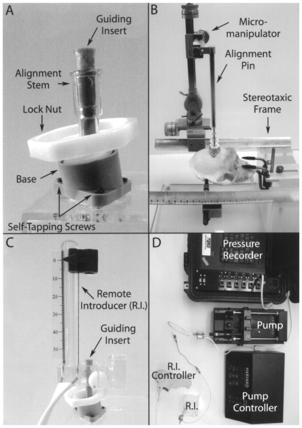
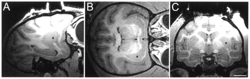
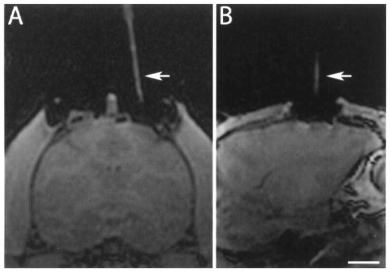
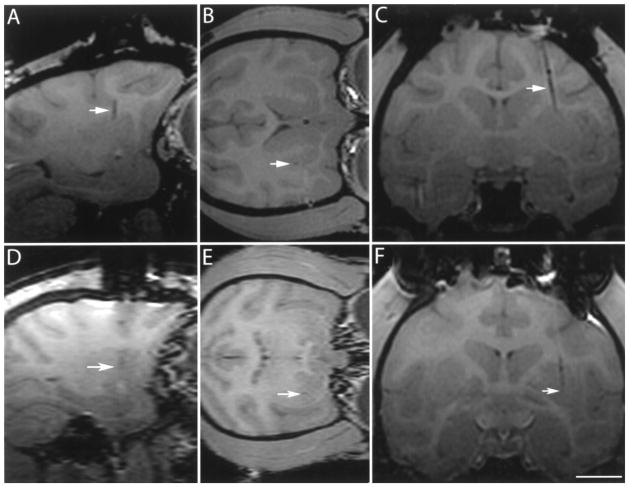
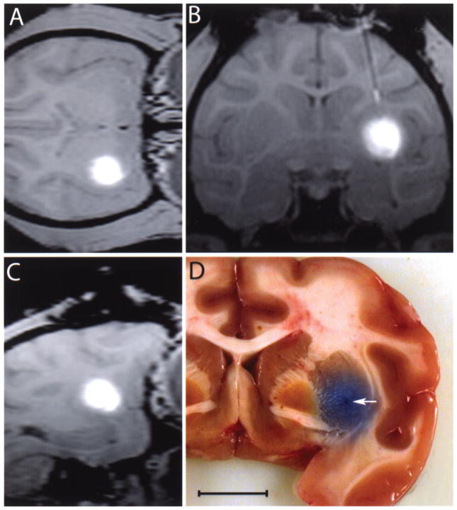
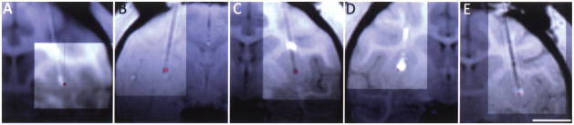
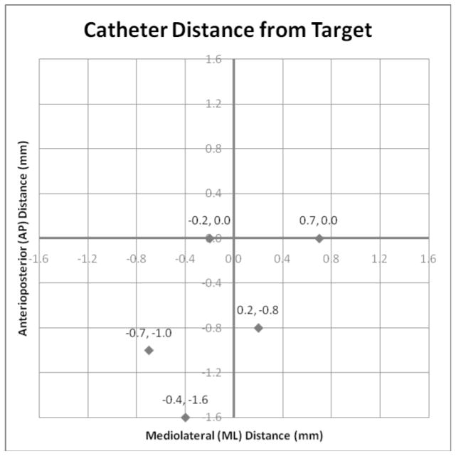
During in vivo intracerebral infusions, the ability to perform accurate targeting towards a 3D-specific point allows control of the anatomical variable and identification of the effects of variations in other factors. Intraoperative MRI navigation systems are currently being used in the clinic, yet their use in nonhuman primates and MRI monitoring of intracerebral infusions has not been reported. In this study rhesus monkeys were placed in a MRI-compatible stereotaxic frame. T1 MRIs in the three planes were obtained in a 3.0T GE scanner to identify the target and plan the trajectory to ventral postcommisural putamen. A craniotomy was performed under sterile surgical conditions at the trajectory entry point. A modified MRI-compatible trajectory guide base (Medtronic Inc.) was secured above the cranial opening and the alignment stem applied. Scans were taken to define the position of the alignment stem. When the projection of the catheter in the three planes matched the desired trajectory to the target, the base was locked in position. A catheter replaced the alignment stem and was slowly introduced to the final target structure. Additional scans were performed to confirm trajectory and during the infusion of a solution of gadoteridol (ProHance, Bracco Diagnostics; 2 mM/L) and bromophenol blue (0.16 mg/ml) in saline. Monitoring of the pressure in the infusion lines was performed using pressure monitoring and infusion pump controller system (Engineering Resources Group Inc.) in combination with a MRI-compatible infusion pump (Harvard). MRI during infusion confirmed successful targeting and matched postmortem visualization of bromophenol blue. Assessment of the accuracy of the targeting revealed an overall 3D mean ± SD distance error of 1.2 ± 0.6 mm and angular distance error of 0.9 ± 0.5 mm. Our results in nonhuman primates confirm the accuracy of intraoperative MRI intracerebral navigation combined with an adaptable, pivot point-based targeting system and validates its use for preclinical intracerebral procedures.
Figures
Similar articles
-
Development of a novel frameless skull-mounted ball-joint guide array for use in image-guided neurosurgery.J Neurosurg. 2019 Feb 15;132(2):595-604. doi: 10.3171/2018.10.JNS182169. Print 2020 Feb 1. J Neurosurg. 2019. PMID: 30771782
-
Remotely-controlled approach for stereotactic neurobiopsy.Comput Aided Surg. 2002;7(4):237-47. doi: 10.1002/igs.10047. Comput Aided Surg. 2002. PMID: 12454894
-
Reliable navigation registration in cranial and spine surgery based on intraoperative computed tomography.Neurosurg Focus. 2019 Dec 1;47(6):E11. doi: 10.3171/2019.8.FOCUS19621. Neurosurg Focus. 2019. PMID: 31786552
-
Stereotactic Biopsy Platforms with Intraoperative Imaging Guidance.Neurosurg Clin N Am. 2017 Oct;28(4):465-475. doi: 10.1016/j.nec.2017.05.002. Neurosurg Clin N Am. 2017. PMID: 28917276 Review.
-
New possibilities for stereotaxis. Information-guided stereotaxis.Stereotact Funct Neurosurg. 2001;76(3-4):159-67. doi: 10.1159/000066714. Stereotact Funct Neurosurg. 2001. PMID: 12378094 Review.
Cited by
-
Feasibility and performance of a frameless stereotactic system for targeting subcortical nuclei in nonhuman primates.J Neurosurg. 2020 Mar 6;134(3):1064-1071. doi: 10.3171/2019.12.JNS192946. Print 2021 Mar 1. J Neurosurg. 2020. PMID: 32114536 Free PMC article.
-
Preclinical evaluation of transaxial intraputaminal trajectory for enhanced distribution of grafted cells in Parkinson's disease.J Neurosurg. 2024 Jul 26;141(6):1554-1566. doi: 10.3171/2024.4.JNS24367. Print 2024 Dec 1. J Neurosurg. 2024. PMID: 39059426 Free PMC article.
-
Using non-invasive neuroimaging to enhance the care, well-being and experimental outcomes of laboratory non-human primates (monkeys).Neuroimage. 2021 Mar;228:117667. doi: 10.1016/j.neuroimage.2020.117667. Epub 2020 Dec 24. Neuroimage. 2021. PMID: 33359353 Free PMC article. Review.
-
Real-Time Intraoperative MRI Intracerebral Delivery of Induced Pluripotent Stem Cell-Derived Neurons.Cell Transplant. 2017 Apr 13;26(4):613-624. doi: 10.3727/096368916X692979. Epub 2016 Sep 14. Cell Transplant. 2017. PMID: 27633706 Free PMC article.
-
SIMULATING CONVECTION-ENHANCED DELIVERY IN THE PUTAMEN USING PROBABILISTIC TRACTOGRAPHY.Proc IEEE Int Symp Biomed Imaging. 2011 Mar-Apr;2011:787-790. doi: 10.1109/ISBI.2011.5872523. Epub 2011 Jun 9. Proc IEEE Int Symp Biomed Imaging. 2011. PMID: 27795809 Free PMC article.
References
-
- Bankiewicz KS, Bringas JR, McLaughlin W, Pivirotto P, Hundal R, Yang B, Emborg ME, Nagy D. Application of gene therapy for Parkinson’s disease: Nonhuman primate experience. Adv Pharmacol. 1998;42:801–806. - PubMed
-
- Bankiewicz KS, Bringas J, Pivirotto P, Kutzscher E, Nagy D, Emborg ME. Technique for bilateral intracranial implantation of cells in monkeys using an automated delivery system. Cell Transplant. 2000;9:595–607. - PubMed
-
- Capitanio JP, Emborg ME. Contributions of non-human primates to neuroscience research. Lancet. 2008;371:1126–1135. - PubMed
-
- Elias WJ, Fu KM, Frysinger RC. Cortical and subcortical brain shift during stereotactic procedures. J Neurosurg. 2007;107:983–988. - PubMed
-
- Emborg ME, Carbon M, Holden JE, During MJ, Ma Y, Tang C, Moirano J, Fitzsimons H, Roitberg BZ, Tuccar E, Roberts A, Kaplitt MG, Eidelberg D. Subthalamic glutamic acid decarboxylase gene therapy: changes in motor function and cortical metabolism. J Cereb Blood Flow Metab. 2007;27:501–509. - PubMed
Publication types
MeSH terms
Grants and funding
LinkOut - more resources
Full Text Sources
Other Literature Sources
Medical
