

"Duplicated" or "multiple" cervical internal carotid and vertebral arteries from fenestration, duplication and vasa vasorum to segmental rete
- PMID: 20587213
- PMCID: PMC3463289
- DOI: 10.1177/159101990401000403
"Duplicated" or "multiple" cervical internal carotid and vertebral arteries from fenestration, duplication and vasa vasorum to segmental rete
Abstract
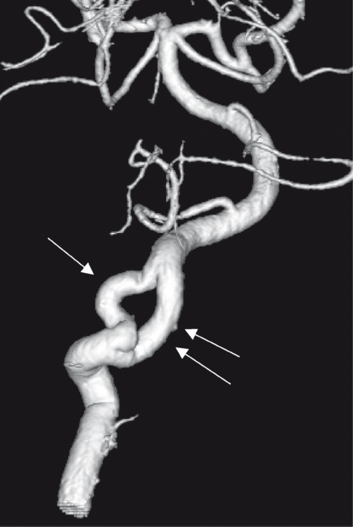
The appearance of multiple cervical arteries may be due to a variety of anatomic situations. Arterial fenestrations and duplications have different anatomic origins, with distinct appearances on angiography. They are associated with incomplete segmental development. The vasa vasorum of the internal carotid artery are rarely seen unless enlarged in pathological situations. They represent a peculiar segmental, in-situ, collateral circulation. Retes, on the other hand, correspond to a more complex reconstitution of an early segmental regression. Careful analysis of each of these features is required to choose the best appropriate terminology. The purpose of this paper is to report illustrative cases to enhance the distinctive features of each disposition.
Figures


Similar articles
-
Revascularization of occluded internal carotid arteries by hypertrophied vasa vasorum: report of four cases.Neurosurgery. 1999 Sep;45(3):634-7. doi: 10.1097/00006123-199909000-00040. Neurosurgery. 1999. PMID: 10493385
-
Carotid pseudostring sign from vasa vasorum collaterals.J Ultrasound Med. 2003 Sep;22(9):959-63; quiz 964-5. doi: 10.7863/jum.2003.22.9.959. J Ultrasound Med. 2003. PMID: 14510268
-
Vasa vasorum of the intracranial arteries.Acta Neurochir (Wien). 1998;140(5):411-6. doi: 10.1007/s007010050118. Acta Neurochir (Wien). 1998. PMID: 9728239
-
[Modeling of elastic deformation and vascular resistance of arterial and venous vasa vasorum].J Mal Vasc. 1998 Oct;23(4):282-8. J Mal Vasc. 1998. PMID: 9827409 Review. French.
-
Arterial fenestrations and their association with cerebral aneurysms.J Clin Neurosci. 2014 Dec;21(12):2184-8. doi: 10.1016/j.jocn.2014.07.005. Epub 2014 Aug 21. J Clin Neurosci. 2014. PMID: 25150765 Review.
Cited by
-
Multichannel internal carotid artery: congenital or acquired pattern?Neurol Sci. 2025 May 24. doi: 10.1007/s10072-025-08252-1. Online ahead of print. Neurol Sci. 2025. PMID: 40411703
-
Revascularization of the internal carotid artery through the hypertrophied vasa vasorum in traumatic carotid-cavernous fistula previously treated by ligation of cervical carotid arteries: A case report.Surg Neurol Int. 2022 Jul 29;13:324. doi: 10.25259/SNI_450_2022. eCollection 2022. Surg Neurol Int. 2022. PMID: 36128096 Free PMC article.
-
[Rete compensation in agenesis of the internal carotid artery].Nervenarzt. 2007 Aug;78(8):948-53. doi: 10.1007/s00115-007-2260-x. Nervenarzt. 2007. PMID: 17457561 German.
-
Double-lumen Carotid Plaque Associated with Severe Stenosis Treated with Staged Angioplasty: A Case Report.NMC Case Rep J. 2021 Jul 3;8(1):359-365. doi: 10.2176/nmccrj.cr.2020-0205. eCollection 2021. NMC Case Rep J. 2021. PMID: 35079489 Free PMC article.
References
-
- Lasjaunias P, Braun JP, et al. True and false fenestration of the vertebral artery. Journal of Neuroradiology. Journal de Neuroradiologie. 1980;7(3):157–166. - PubMed
-
- Lasjaunias P, Berenstein A, TerBrugge KG. Surgical Neurorangiography. 2nd edition. Vol 1. Berlin Heidelberg New York: Springer-Verlag; 2001. pp. 590–596.
-
- Takaba M, Endo S, et al. Vasa vasorum of the intracranial arteries. Acta Neurochir. 1998;140(5):411–416. - PubMed
-
- Williams JK, Heistad DD. The vasa vasorum of the arteries. Journal des Maladies Vascularies. 1996;21(Sup C):266–269. - PubMed
-
- Berenstein A, Lasjaunias P, TerBrugge KG. Surgical Neurorangiography. 2nd edition. Vol 2. Berlin Heidelberg New York: Springer-Verlag; 2004. pp. 314–315. 978-979.
LinkOut - more resources
Full Text Sources
