

doi: 10.1177/159101990401000408.
Epub 2005 Feb 8.
Multiple cerebral aneurysms in a patient with recurrent cardiac myxomas. A case report
Affiliations
- PMID: 20587218
- PMCID: PMC3463294
- DOI: 10.1177/159101990401000408
Item in Clipboard
Multiple cerebral aneurysms in a patient with recurrent cardiac myxomas. A case report
Interv Neuroradiol.
.
Abstract
A 22 year old female presented in 1987 a cardiac myxoma, removed in 1991. In 1992 catheter angiography showed bilateral aneurysms. Conservative treatment was elected. In 2000 recurrence of the myxoma and subsequent removal, prompted new angiography. All aneurysms had decreased in size, some had spontaneously disappeared.
Figures
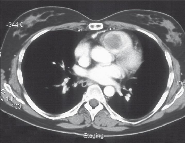
CT scan demonstrates right ventricular mass (myxoma).
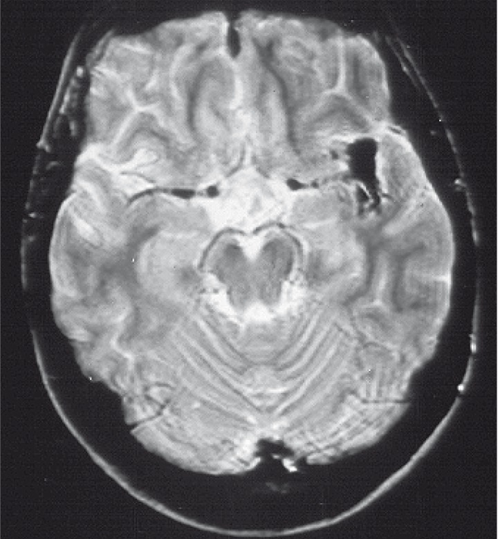
Axial T2w image demonstrates unusual shape of trifurcation of left MCA.
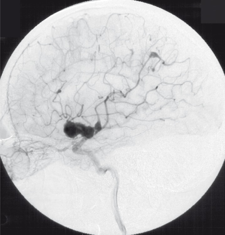
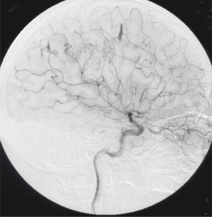
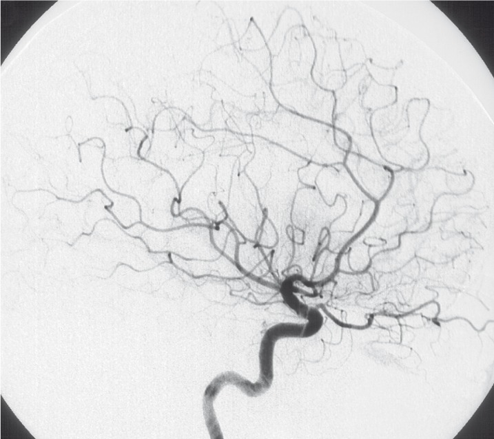
Injection of left ICA lateral view (1992).
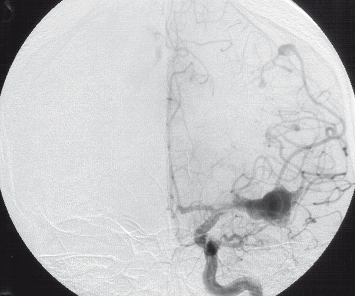
Injection of left ICA frontal view (1992).
Injection of right ICA lateral view (1992).
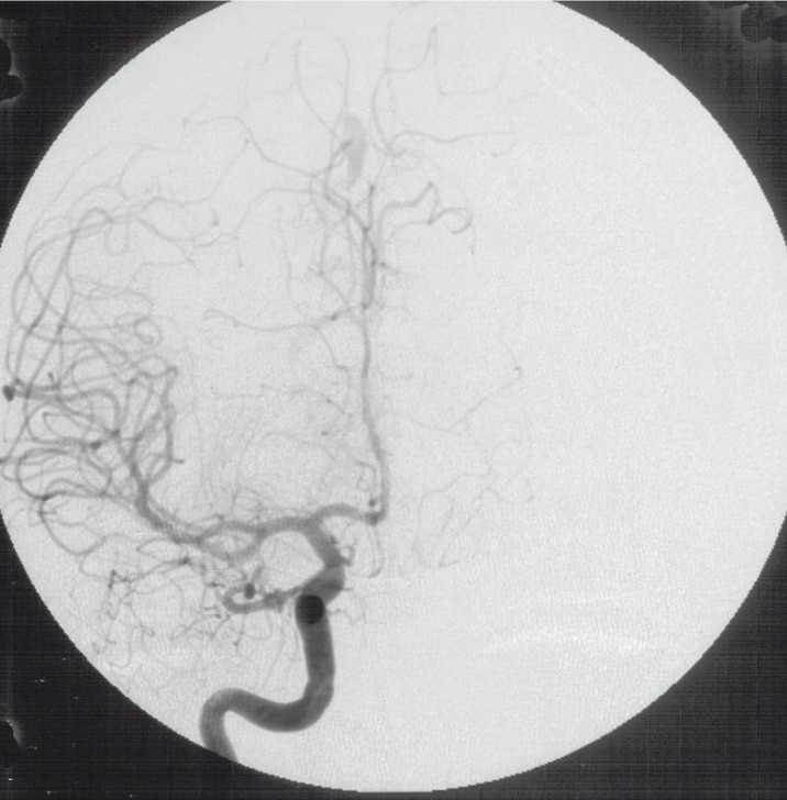
Injection of right ICA frontal view (1992).
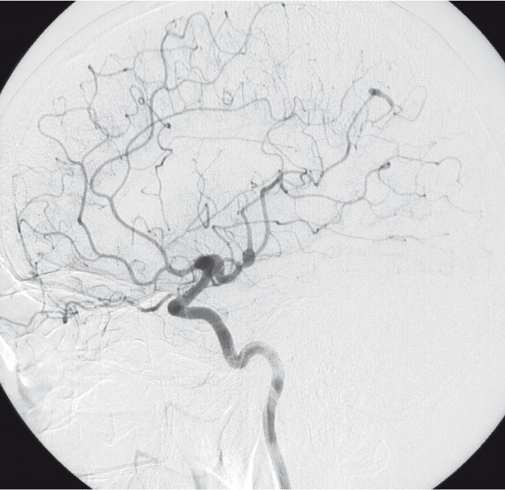
Injection of left ICA lateral view (2003).
Injection of left ICA frontal view (2003).
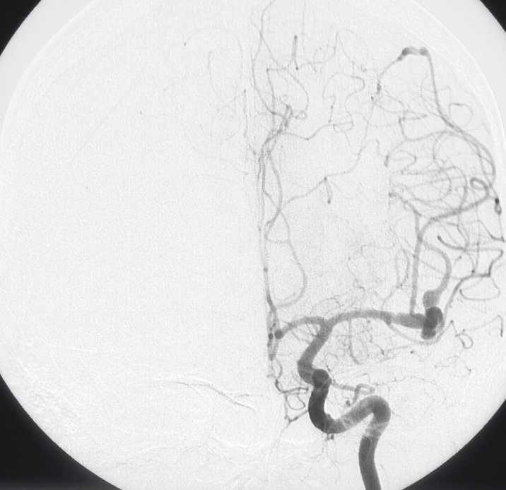
Injection of right ICA lateral view (2003).
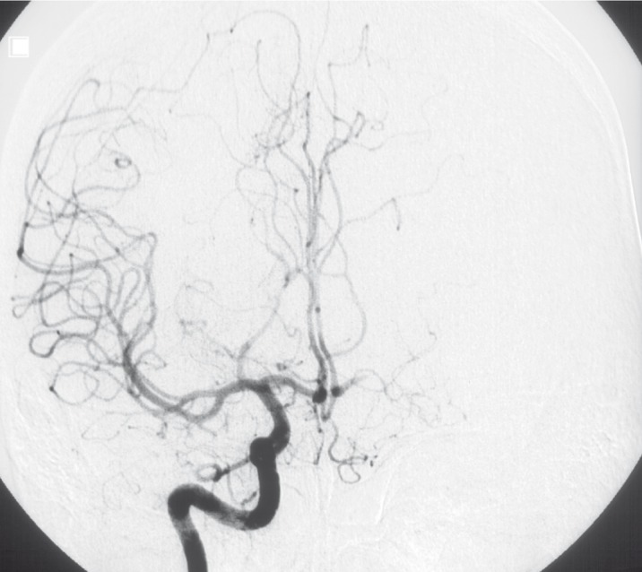
Injection of right ICA frontal view (2003).
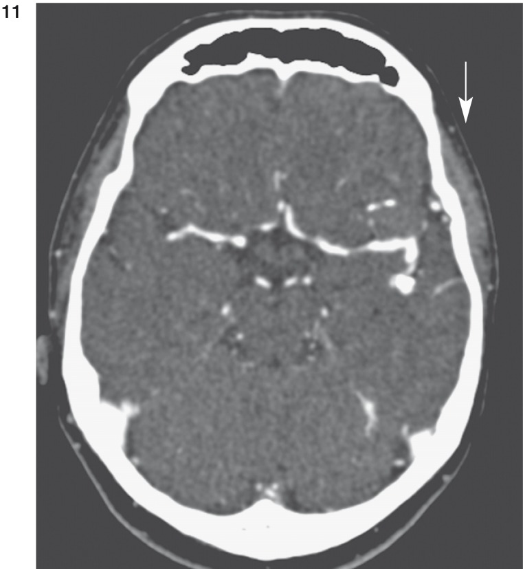
CT scans demonstrate marginal calcification (arrow in fig. 11) and thrombosis (arrow in fig. 12) of large aneurysm of left MCA.
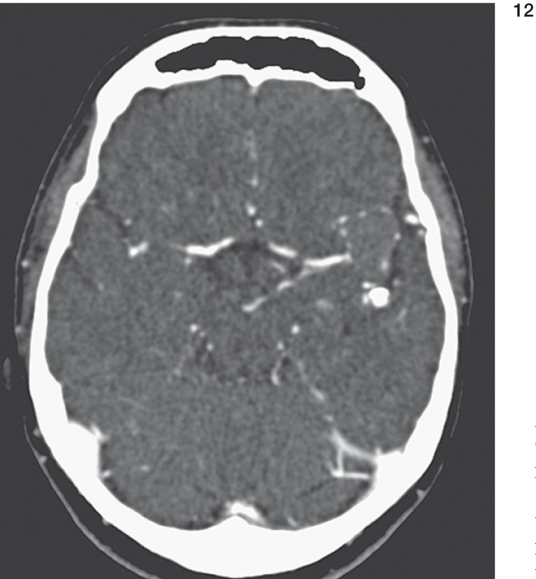
CT scans demonstrate marginal calcification (arrow in fig. 11) and thrombosis (arrow in fig. 12) of large aneurysm of left MCA.
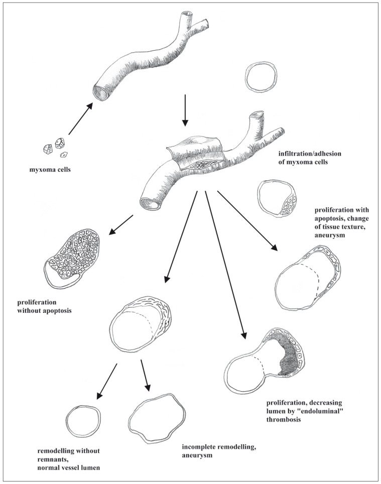
Pathogenesis of vessel involvement in myxoma.
Similar articles
-
The dynamic natural history of cerebral aneurysms from cardiac myxomas: A review of the natural history of myxomatous aneurysms.Interv Neuroradiol. 2018 Jun;24(3):277-283. doi: 10.1177/1591019917754070. Epub 2018 Jan 31. Interv Neuroradiol. 2018. PMID: 29383976 Free PMC article. Review.
-
Multiple cerebral aneurysms as delayed complication of left cardiac myxoma: a case report and review.Acta Neurol Scand. 2005 Jun;111(6):345-50. doi: 10.1111/j.1600-0404.2005.00413.x. Acta Neurol Scand. 2005. PMID: 15876333 Review.
-
Late Diagnosis of Multiple Cerebral Aneurysms A Decade after Resection of Cardiac Myxoma.Case Rep Neurol. 2020 Dec 14;12(Suppl 1):189-195. doi: 10.1159/000505390. eCollection 2020 Sep-Dec. Case Rep Neurol. 2020. PMID: 33505294 Free PMC article.
-
Multiple cerebral and coronary aneurysms in a patient with left atrial myxoma.Int J Cardiovasc Imaging. 2012 Dec;28 Suppl 2:129-32. doi: 10.1007/s10554-012-0140-3. Epub 2012 Oct 25. Int J Cardiovasc Imaging. 2012. PMID: 23097127
-
Multiple intracranial aneurysms as delayed complications of an atrial myxoma: case report.Neurosurgery. 2001 Jul;49(1):200-2; discussion 202-3. doi: 10.1097/00006123-200107000-00031. Neurosurgery. 2001. PMID: 11440443
Cited by
-
Anesthesia management of atrial myxoma resection with multiple cerebral aneurysms: a case report and review of the literature.BMC Anesthesiol. 2020 Jul 4;20(1):164. doi: 10.1186/s12871-020-01055-1. BMC Anesthesiol. 2020. PMID: 32622360 Free PMC article. Review.
-
The role of lipids in corneal diseases and dystrophies: a systematic review.Clin Transl Med. 2017 Dec;6(1):30. doi: 10.1186/s40169-017-0158-1. Epub 2017 Aug 11. Clin Transl Med. 2017. PMID: 28782089 Free PMC article. Review.
-
Delayed multiple intracranial aneurysms caused by left atrial myxoma: a case report and literature review.Transl Pediatr. 2022 Jan;11(1):149-156. doi: 10.21037/tp-21-11. Transl Pediatr. 2022. PMID: 35242661 Free PMC article.
-
Cerebral Aneurysms Caused by Atrial Myxoma-A Systematic Review of the Literature.J Pers Med. 2022 Dec 21;13(1):8. doi: 10.3390/jpm13010008. J Pers Med. 2022. PMID: 36675669 Free PMC article. Review.
-
The dynamic natural history of cerebral aneurysms from cardiac myxomas: A review of the natural history of myxomatous aneurysms.Interv Neuroradiol. 2018 Jun;24(3):277-283. doi: 10.1177/1591019917754070. Epub 2018 Jan 31. Interv Neuroradiol. 2018. PMID: 29383976 Free PMC article. Review.
References
-
- Kazuhide F, Sasaki T, et al. Histologically verified cerebral aneurysm formation secondary to embolism from cardiac myxoma. J Neurosurg. 1995;83:170–173. - PubMed
-
- Stoane L, Allan JH, Jr, Collins HA. Radiologic observations in cerebral embolization from left heart myxomas. Radiology. 1966;87:262–266. - PubMed
-
- New PFJ, Price DL, Carter B. Cerebral angiography in cardiac myxoma. Correlation of angiographic and histopathological findings. Radiology. 1970;96:335–345. - PubMed
-
- Roeltgen DP, Weimer GR, Patterson LF. Delayed neurologic complications of left atrial myxoma. Neurology. 1981;31:8–13. - PubMed
-
- Mattle HP, Maurer D, et al. Schroth Cardiac myxomas: a long term study. J Neurol. 1995;242(10):689–694. - PubMed
LinkOut - more resources
Full Text Sources
