. 2004 Mar 30;10 Suppl 1(Suppl 1):85-92.
doi: 10.1177/15910199040100S113. Epub 2008 Jun 9.

Transvenous embolization of dural arteriovenous fistula of the cavernous sinus. Fistulous points and route of catheterization

Affiliations

Transvenous embolization of dural arteriovenous fistula of the cavernous sinus. Fistulous points and route of catheterization

S Takahashi et al. Interv Neuroradiol. .

Abstract

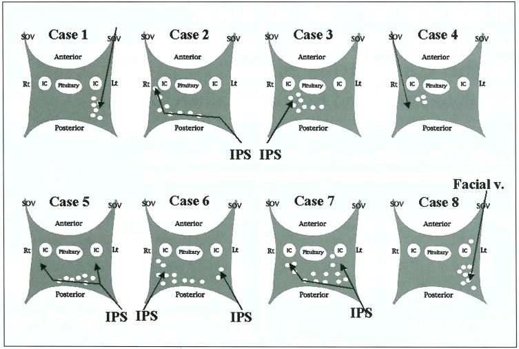
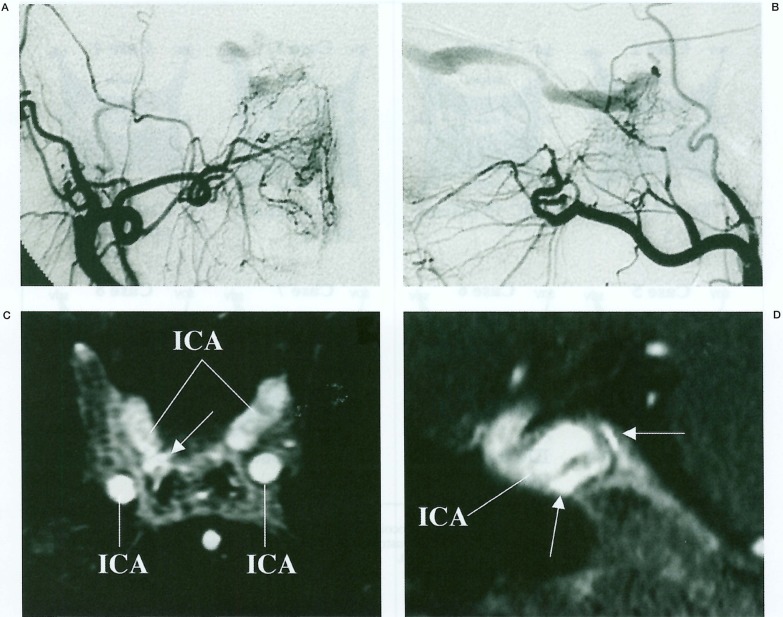
We reviewed magnetic resonance (MR) images and digital subtraction angiograms (DSA) from eight patients with dural arteriovenous fistula of the cavernous sinus (DAVFCS) to clarify the fistulous points and to evaluate the venous access routes into the cavernous sinus for transvenous embolization (TVE). Multiplanar reconstruction of the MR images was achieved using three-dimensional fast spoiled gradient-recalled acquisition in the steady state (3-D fast SPGR) after the intravenous administration of gadopentetate dimeglumine (Gd-DTPA). TVE was performed using microcoils via the inferior petrosal sinus (IPS) using the transfemoral approach in five patients, via the facial vein and superior ophthalmic vein (SOV) using the transfemoral approach in 1 patient, and by SOV puncture in two patients. Most fistulas were detected in the posterior portion of the cavernous sinus or in the posterior intercavernous sinus in all of the patients. Fistulas identified as hyperintense dots or lines on contrast-enhanced 3-D fast SPGR images and were replaced with the microcoils. Target embolization of the fistulas was feasible in three patients treated via the SOV and in one patient treated via the IPS. Contrast- enhanced 3-D fast SPGR can help to identify the fistulous points of DAVFCS. Precise identification of fistulous points and selection of the adequate access route are mandatory for efficient TVE of DAVFCS.


Figures

Figure 1
Figure 1
Cavernous sinus: fistulous points and venous access routes. Fistulous points are shown as white dots in the cavernous sinus from axial source images obtained by contrast-enhanced 3-D fast SPGR. Black arrows reveal venous access routes in TVE. IC, internal carotid artery; pituitary, pituitary gland; Rt, right; Lt, left; SOV, superior ophthalmic vein; IPS, inferior petrosal sinus; Facial v., facial vein.
Figure 2
Figure 2
Microcoils positioned in the cavernous sinus. Microcoils placed in the cavernous sinus are illustrated as black tangles. Numbers of IDCs and FPCs are described below the illustration. Numbers in parentheses are total lengths of IDCs. IDC, interlocking detachable coil; FPC, fibered platinum coil.
Figure 3
Figure 3
Case 4. Right external carotid arteriogram, frontal (A) and lateral (B) projections, shows DAVF of the right cavernous sinus with venous drainage into the right superior ophthalmic vein. The axial source image (C) and reconstructed sagittal image (D) obtained by contrast-enhanced 3-D fast SPGR show the point-shaped or linear hyperintensity (arrows) abutting the right internal carotid artery in the right cavernous sinus. ICA, internal carotid artery.
Figure 4
Figure 4
Case 4. Right external carotid arteriogram, fronal (A) and lateral (B) projections, immediately after TVE using microcoils (arrows) shows disappearance of DAVF. The axial source image (C) and reconstructed sagittal image (D) obtained by contrast-enhanced 3-D fast SPGR show the microcoils placed on the fistulous points (arrows).
Figure 5
Figure 5
Case 6. Left common carotid arteriogram, frontal (A) and lateral (B) projections, show DAVFCS draining into the bilateral IPS. The axial source image of the cavernous sinus (C) and the reconstructed coronal image of the posterior intercavernous sinus (D) obtained by contrast-enhanced 3-D fast SPGR show the multiple hyperintense curvilinear structures (arrows) posteriorly in bilateral cavenous sinuses and the posterior intercavernous sinus. Left internal carotid arteriogram, frontal view (E), during coil packing of the right cavernous sinus shows residual DAVF with venous drainage into the right SOV diverted from the right IPS. Right SOV injection, basal view (F), after the navigation of the microcatheter through the placed microcoils shows the residual space of the right cavernous sinus and right SOV. ICA, internal carotid artery.
Figure 6
Figure 6
Case 6. Right common carotid arteriogram, fronal (A) and lateral (B) projections, immediately after TVE using microcoils (arrows) shows disappearance of DAVE The axial source image (C) and reconstructed coronal image (D) obtained by contrast-enhanced 3-D fast SPGR show the microcoils (arrows) placed in bilateral cavernous sinuses and the posterior intercavernous sinus. ICA, internal carotid artery.

Similar articles

Cited by

References

    1. Teng MMH, Guo WY, et al. Occlusion of arteriovenous malformations of the cavernous sinus via the superior ophthalmic vein. Am J Neuroradiol. 1988;9:539–546. - PMC - PubMed
    1. Monsein LH, Debrun GN, et al. Treatment of dural carotid-cavernous fistulas via the superior ophthalmic vein. Am J Neuroradiol. 1991;12:435–439. - PMC - PubMed
    1. Halbach VV, Higashida RT, et al. Transvenous embolization of dural fistulas involving the cavernous sinus. Am J Neuroradiol. 1989;10:377–383. - PMC - PubMed
    1. Yamashita K, Taki W, et al. Transvenous embolization of dural caroticocavernous fistulae: technical condiderations. Neuroradiology. 1993;35:475–479. - PubMed
    1. Benndorf G, Bender A, et al. Transvenous occlusion of dural cavernous sinus fistulas through the thrombosed inferior petrosal sinus: report of four cases and review of the literature. Surg Neurol. 2000;54:42–54. - PubMed

LinkOut - more resources