. 2004 Mar 30;10 Suppl 1(Suppl 1):127-34.
doi: 10.1177/15910199040100S122. Epub 2008 Jun 9.

Study of Dural Arteriovenous Fistula Drains into Leptomeningeal Vein without Sinus Interposition

Affiliations

Study of Dural Arteriovenous Fistula Drains into Leptomeningeal Vein without Sinus Interposition

T Kawaguchi et al. Interv Neuroradiol. .

Abstract

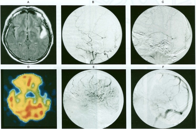
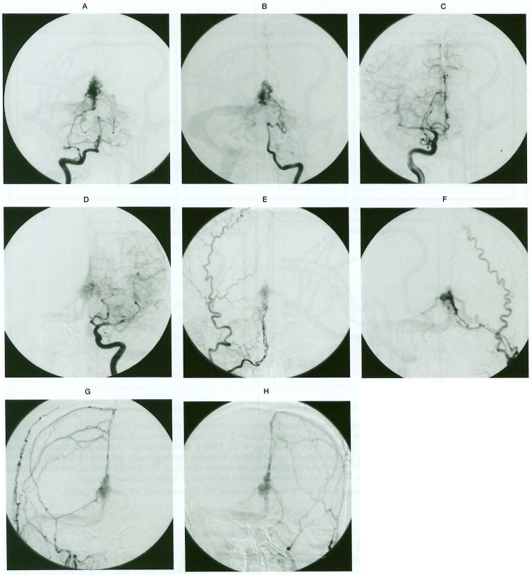
We evaluated dural arteriovenous fistulas (DAVF) drains into leptomeningeal vein (LMV) without the venous sinus interposition. This type of DAVF contained the extra-sinusal type DAVF and the DAVF with so-called pure leptomeningeal venous drainage (PLMVD).We studied 15 patients with DAVF that flows into LMVD without passing into the sinus. The subjects were 5 patients with DAVF in the anterior cranial fossa, 2 with DAVF in the tentorium cerebelli, and 3 with DAVF in the craniocervical junction as extra-sinusal type DAVF and 3 with DAVF in the transverse sigmoid sinus and 2 with DAVF in the superior sagittal sinus as DAVF with PLMVD. This type appears to take a very aggressive course. The arterial pressure of the shunt is directly applied to LMV, which causes bending and winding of the vein, eventually varices, inducing intracranial haemorrhage or venous ischemia in the LMV reflux area. Emergency treatment should be performed as soon as possible. Although it is recognized that interruption of the draining vein is very effective, treatment methods such as TAE, direct surgery, and g knife treatment, or their combinations should be carefully chosen for each case.


Figures

Figure 1
Figure 1
The conception of DAVF was illustrated. This study focused on extrasinusal DAVF and DAVF with PLMVD.
Figure 2
Figure 2
A-C, E-G) Right internal carotid angiograms demonstrating DAVF at the anterior cranial fossa fed by the anterior ethmoidal artery. Main drainages were olfactory vein into basal vein, straight sinus, left transverse-sigmoid sinus. D) 123I-IMP SPECT showing a low perfusion area in the left occipital region. H) Scheme demonstrating the DAVF in the right anterior cranial fossa. Main drainage was olfactory vein into basal vein, straight sinus, left transverse-sigmoid sinus. Normal venous circulation of superior sagittal sinus drain into the right transverse-sigmoid sinus.
Figure 3
Figure 3
A) FLAIR image showed a high sinal intensity area in the left temporal lobe. B, C) Left external carotid angiograms showing the DAVF of the transverse-sigmoid sinus fed by occipital, middle meningeal arteries. The draining vein was vein of Labbé. D) 123I-IMP SPECT showing a low perfusion area in the left temporal region. E,F) Left internal carotid angiograms demonstrating venous congestion in the left temporal lobe. Left transverse and sigmoid sinuses were patent at the venous phase.
Figure 4
Figure 4
A) CT scans demonstrated a subcortical haemorrhage in the right occipital lobe. B) Right external carotid angiogram, lateral view. Right external carotid angiogram revealing the DAVF in the transverse sigmoid sinus. The feeding vessels were middle meningeal artery, occipital artery. C) Scheme demonstrating the transverse sigmoid sinus DAVF with pure leptomenigeal venous drainage, varix. D) Right internal carotid angiogram, lateral view. Right internal carotid angiogram demonstrating that transvese sigmoid sinus was patent.
Figure 5
Figure 5
A) CT scan showing haemorrhage in the fourth ventricle. B) Rt vertebral angiograms showing DAVF and venous aneurysm at the craniocervical junction. An unruptured basilar midportion aneurysm was observed. C) Operative photograph demonstrated CCJ-DAVF with venous aneurysm. D) Scheme of CCJ-DAVF. Feeding vessels were C1 and C2 radicular artery. Draining vessels were posterior medullary vein with venous aneurysm and anterior medullary vein.
Figure 6
Figure 6
A) Right vertebral angiogram, AP view. B) Left vertebral angiogram, AP view. C) Right internal carotid angiogram, AP view. D) Left internal carotid angiogram, AP view. E) Right selective occipital angiogram, AP view. F) Left selective occipital angiogram, AP view. G) Right external carotid angiogram, AP view. H) Left external carotid angiogram, AP view. The DAVF of the tentorium and a round dilatation of a vein of Galen was revealed fed by multiple arteries. Venous drainages were a vein of Galen, straight sinus, and bilateral basal veins.
Figure 7
Figure 7
Illustration showing the modified Lalwani’s classification of transverse-sigmoid sinus DAVF (TS DAVF). A) Group 1. TS DAVF without restriction of parent sinus. B) Group 2. TS DAVF with stenosis of parent sinus. C) Group 3. TS DAVF with one side occlusion(thrombosis) of parent sinus. D) Group 4. TS DAVF with proximal and distal portion occlusion (thrombosis) of parent sinus. This group has retrograde leptomeningeal venous drainage only and was called as TS DAVF with isolated sinus. E) Group 5. TS DAVF with pure leptomeningeal venous drainage. The parent sinus is patent. Subgroup was the presence of LMVD, the presence of venous aneurysm and the presence of spinal venous drainage. OA: occipital artery, MMA: middle meningeal artery, TS: transverse sinus, SS: sigmoid sinus, LV: vein of Labbé.

References

    1. Lasjaunias P, Chiu M, et al. Neurological manifestations of intracranial dural arteriovenous malformations. J Nuerosurg. 1986;64:724–730. - PubMed
    1. Borden JA, Wu JK, et al. A proposed classification for spinal and cranial dural arteriovenous fistulous malformations and implications for treatment. J Neurosurg. 1995;82:166–179. - PubMed
    1. Cognard C, Gobin YP, et al. Cerebral dural arteriovenous fistulas: Clinical and angiographic correlation with a revised classification of venous drainage. Radiology. 1995;194:671–680. - PubMed
    1. Collice M, D’Aliberti G, et al. Surgical interruption of leptomeningeal drainage as treatment for intracranial dural arteriovenous fistulas without dural sinus drainage. J Neurosurg. 1996;84:810–817. - PubMed
    1. Collice M, D’Aliberti G, et al. Surgical treatment of intracranial dural arteriovenous fistulae: role of venous drainage. Neurosurgery. 2000;47:56–67. - PubMed

LinkOut - more resources