

Modeling stromal-epithelial interactions in disease progression
- PMID: 20587339
- PMCID: PMC4195242
Modeling stromal-epithelial interactions in disease progression
Abstract
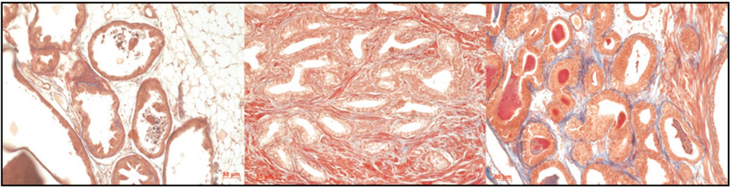
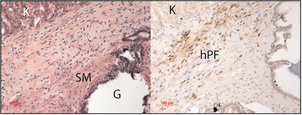
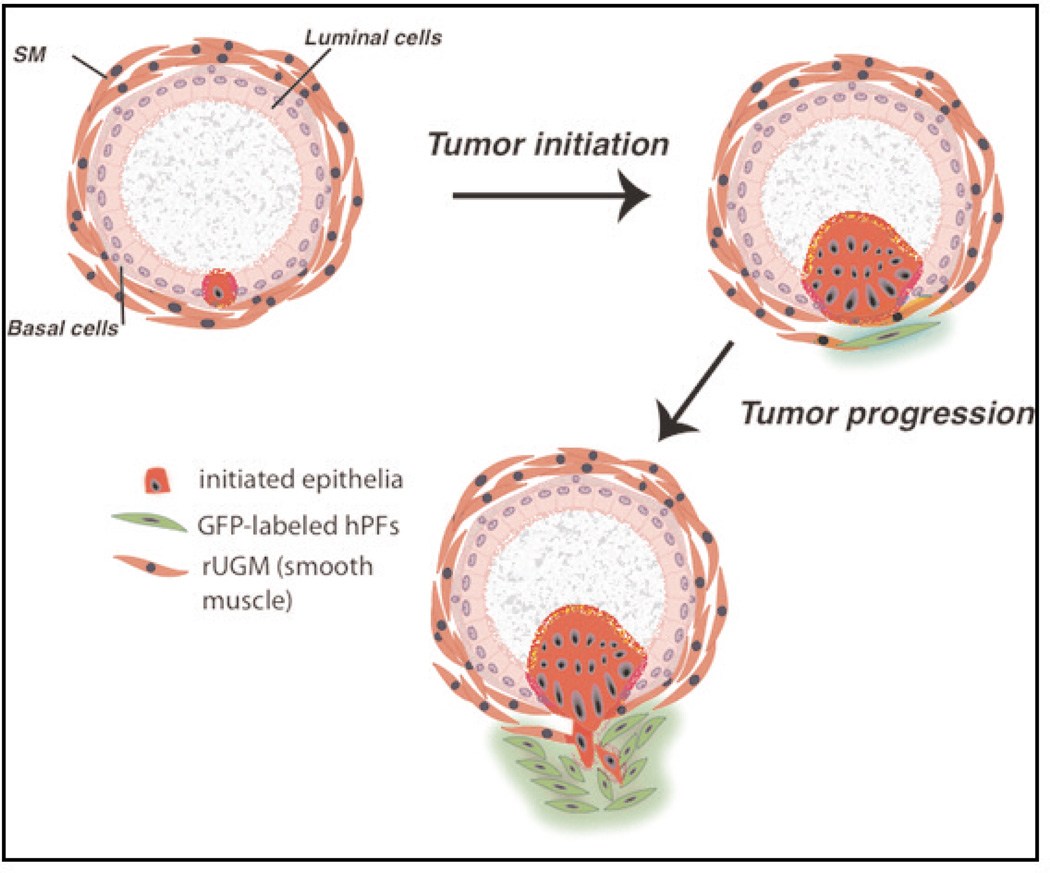
The role of tumor stroma in progression to malignancy has become the subject of intense experimental and clinical interest. The stromal compartment of organs is composed of all the non-epithelial cell types and maintains the proper architecture and nutrient levels required for epithelial and, ultimately, organ function. The composition of the reactive stroma surrounding tumors is vastly different from normal stromal tissue. Stromal phenotype can be correlated with, and predictive of, disease recurrence. In addition, the stroma is now seen as a legitimate target for therapeutic intervention. Although much has been learned about the role of the stromal compartment in development and disease in recent years, a number of key questions remain. Here we review how some of these questions are beginning to be addressed using new models of stromal-epithelial interaction.
Figures
References
-
- Anderson AR, Weaver AM, Cummings PT, Quaranta V. Tumor morphology and phenotypic evolution driven by selective pressure from the microenvironment. Cell. 2006;127(5):905–915. - PubMed
-
- Anderson ARA, Chaplain MAJ, Newman EL, Steele RJC, Thompson AM. Mathematical modelling of tumour invasion and metastasis. Comput Math Methods Med. 2000;2(2):129–154.
-
- Ao M, Franco OE, Park D, Raman D, Williams K, Hayward SW. Cross-talk between paracrineacting cytokine and chemokine pathways promotes malignancy in benign human prostatic epithelium. Cancer Res. 2007;67(9):4244–4253. - PubMed
Publication types
MeSH terms
Grants and funding
LinkOut - more resources
Full Text Sources