. 2010 Nov;38(20):6930-42.
doi: 10.1093/nar/gkq465. Epub 2010 Jun 28.

When simple sequence comparison fails: the cryptic case of the shared domains of the bacterial replication initiation proteins DnaB and DnaD

Affiliations

When simple sequence comparison fails: the cryptic case of the shared domains of the bacterial replication initiation proteins DnaB and DnaD

Farhat Y Marston et al. Nucleic Acids Res. 2010 Nov.

Abstract

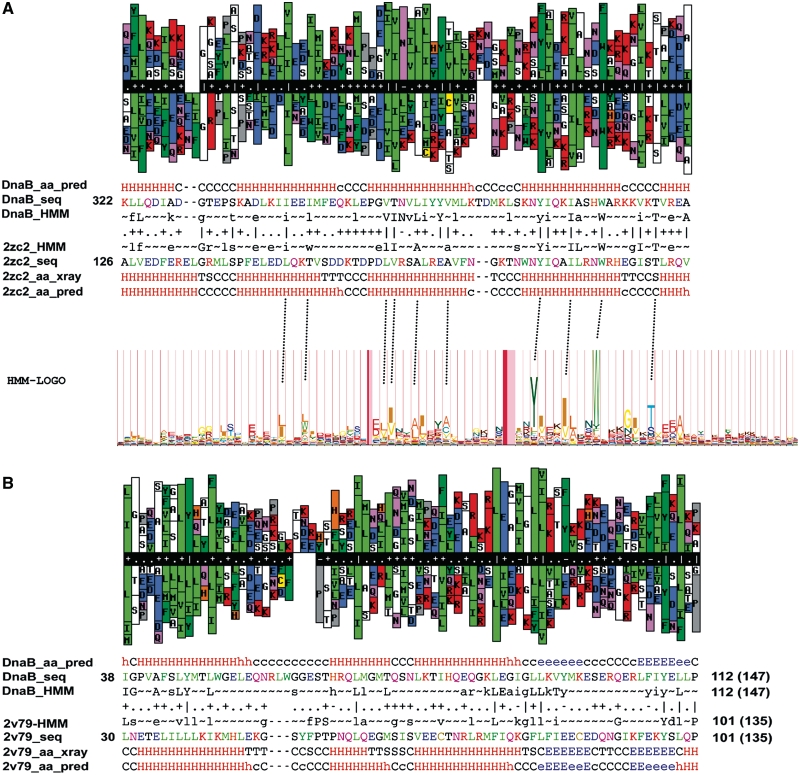
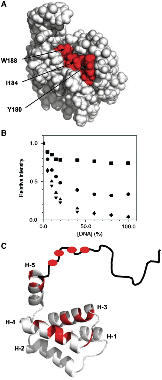
DnaD and DnaB are essential DNA-replication-initiation proteins in low-G+C content Gram-positive bacteria. Here we use sensitive Hidden Markov Model-based techniques to show that the DnaB and DnaD proteins share a common structure that is evident across all their structural domains, termed DDBH1 and DDBH2 (DnaD DnaB Homology 1 and 2). Despite strong sequence divergence, many of the DNA-binding and oligomerization properties of these domains have been conserved. Although eluding simple sequence comparisons, the DDBH2 domains share the only strong sequence motif; an extremely highly conserved YxxxIxxxW sequence that contributes to DNA binding. Sequence alignments of DnaD alone fail to identify another key part of the DNA-binding module, since it includes a poorly conserved sequence, a solvent-exposed and somewhat unstable helix and a mobile segment. We show by NMR, in vitro mutagenesis and in vivo complementation experiments that the DNA-binding module of Bacillus subtilis DnaD comprises the YxxxIxxxW motif, the unstable helix and a portion of the mobile region, the latter two being essential for viability. These structural insights lead us to a re-evaluation of the oligomerization and DNA-binding properties of the DnaD and DnaB proteins.


Figures

Figure 1.
Figure 1.
Defining the extent of the C-terminal domain of DnaD. (A) NMR 15N relaxation (T2) data, which defines regions of mobility in solution. The backbone from residue 129 to 206 shows uniform T2 values, with elevated values beyond residue 206 indicating that the boundary between structure and disorder is approximately at residue 206. The reduced value of T2 for the four data points shown at the C-terminus suggest some degree of constraint or conformational broadening, but there is no evidence for regular structure in the NOEs, chemical shifts or DNA-binding intensity changes for these residues. An alignment of B. subtilis DnaD-Cd with the sequence from S. mutans, corresponding to the structure 2zc2 is also shown. The extent of helices in the two structures is shown in grey above and below the sequences. Identical residues are coloured red, and similar residues are colored yellow. (B) ClustalW alignment of DnaD sequences. The sequences are sequences 1, 6, 11, 16, etc. drawn from the collection in Supplementary Figure S3. Residues that are identical in at least 50% of sequences (excluding gaps) are colored red. Residues that are similar across at least 50% of sequences are colored yellow. Sequences are marked with their Uniprot identifier. Below the alignment the fraction, fNG, of basic (blue) and acidic (red) residues is shown, expressed as a fraction of the total non-gapped residues at each position in the alignment in Supplementary Figure S3. The profile was smoothed using a three-residue window. Across all parts of this figure the data are aligned vertically by residue number of B. subtilis DnaD, except where extensive gapping prevents this beyond residue 215.
Figure 2.
Figure 2.
Domain structure of DnaD and DnaB. The figure shows the experimentally determined secondary structure of DnaD and the predicted secondary structure of DnaB (Supplementary Figure S1). The secondary-structure elements are color coded to show the regions for which HHpred detects homology. The estimated extent of the DDBH1 and DDBH2 domains in the two proteins is shown.
Figure 3.
Figure 3.
HMM relationships between DnaD and DnaB. (A) Top HHpred PDB hit obtained with a query of residues 306–472 of DnaB. The hit is the 2zc2 structure which is closely homologous to the B. subtilis DnaD-Cd. The HMM-LOGO for the DDBH2 domain of the combined sequence set of 326 sequences is shown below the HHpred alignment, with the correspondence of the highest probability residue types between the two HMMs shown by dashed lines. (B) Top HHpred PDB hit obtained with a query of residues 1–147 of DnaB. The hit is the 2v79 X-ray structure of the B. subtilis DnaD-Nd domain. In the HHpred, alignment parts of (A) and (B) the following abbreviations are used: ss_pred, secondary-structure prediction by PSIPRED; seq, sequence; HMM, HHpred representation of HMM where upper and lower case letters denote amino acids with >60 and >40% probability, respectively. Above the alignment are histograms of the DnaB and the PDB HMMs. Bars are proportional to probability of a certain amino acid type at each position, with bars omitted for probabilities <10%. The alignments are images returned by HHpred. The HMM-LOGO was created using the logomat-M server (http://www.sanger.ac.uk/cgi-bin/software/analysis/logomat-m.cgi).
Figure 4.
Figure 4.
The conserved YxxxIxxxW motif and DNA-binding data mapped onto a structural model of DnaD-Cd. (A) A space filling representation of the DnaD-Cd model, with the six most highly conserved residues in DnaD-Cd (A166, A170, Y180, I184, L185 and W188) colored red. Three of these residues (W188, Y180 and I184) form an exposed hydrophobic patch, whereas A166, A170 and L185 appear to anchor this segment to the main hydrophobic core of the protein. (B) Examples of three types of behavior observed on titration of 10 mer DNA into DnaD-Cd. HSQC crosspeaks for the majority of residues (such as E134, circles) show a moderate intensity reduction to ∼30% of initial value. Some residues show a marked intensity reduction (residue I184, triangles; residue K205, inverted triangles). Some residues (residue W229, squares) show only a very small reduction in intensity. DNA concentration is expressed as fraction of protein concentration. Data for all residues are presented in Supplementary Figure S6. (C) Localization of DNA binding by perturbation of 15N HSQC crosspeaks. The structure is shown in cartoon representation in the same orientation as (A), with the C-terminal end of helices labeled. The mobile portion is depicted in sketch form. Residues for which the crosspeak intensity is reduced by >90% after addition of 1 : 1 10-mer DNA to DnaD-Cd215 are colored red. These are residues 168, 170, 171, 176, 177, 181, 183, 184, 185, 188, 201, 203, 205, 207, 208, 210 and 211. This figure is based on data for DnaD-Cd215 sample since it gave superior HSQC spectra (presumably due to superior tumbling properties). A closely similar subset of residues is selected with an 80% cutoff in the titration with DnaD-Cd. The slightly greater drop in intensity in the DnaD-Cd215 titration probably arises from the increased sharpness of the lines for the apo-protein in this construct.
Figure 5.
Figure 5.
DNA binding of DnaD-Cd and truncation mutants. (A) Y180A and W188A have reduced DNA-binding activity. Agarose gel shift assays using DnaD, Y180A and W188A complexes and supercoiled pBSK plasmid. Comparative experiments were carried out with increasing concentrations of proteins, as indicated. Both mutant proteins were defective in DNA binding compared to wild-type DnaD. (B) Direct comparison of the DNA-binding activities of full length DnaD and the mutant Y180A, W188A proteins. Gel shift assays were carried out in 100 mM NaCl, 50 mM Tris pH 7.5, 1 mM EDTA, 1 mM DTT, 2.2% v/v glycerol with 0.625 nM ssDNA or 0.125 nM dsDNA probes at increasing concentrations of proteins, as indicated. (C) DnaD-Cd196 is deficient in DNA binding. DNA-binding reactions were carried out with increasing concentrations of DnaD and DnaD-Cd196 proteins as indicated in 30 mM NaCl, 50 mM Tris pH 7.5, 1 mM EDTA, 1 mM DTT and 0.625 nM ss or dsDNA probes, as indicated. (D) DnaD-Cd215 binds DNA but DnaD-Cd206 does not. DNA-binding reactions were carried out with increasing concentrations of DnaD-Cd206 and DnaD-Cd215 proteins as in described in panel (C).
Figure 6.
Figure 6.
Biological significance of DnaD binding to DNA. (A) Truncated DnaD-V196-STOP does not rescue growth of dnaD23ts at the non-permissive temperature. The dnaD23ts strains carrying an ectopic inducible wild-type dnaD (WKS810), dnaDY180A (WKS812), dnaDW188A (WKS816) or dnaD-V196-STOP (WKS818) were grown on LB agar. A dnaD23ts strain without inducible dnaD (KPL73) is shown as control. Plates were incubated overnight at 30°C or 48°C as indicated. (B) DnaD-Y180A and DnaD-W188A sustain growth. Strains carrying an inducible wild-type DnaD (WKS876), Y180A (WKS878), W188A (WKS880) and DnaD23 (WKS883) can support growth at 30°C when the native copy of dnaD is knocked out. LB agar plates were incubated over night at the indicated temperature. As a control, 48°C is shown for WKS883. (C) Overproduction of wild-type DnaD causes growth inhibition. Strains were grown on LB agar plates, without or with 1-mM IPTG over night at 37°C. WKS802-808 contain an ectopic inducible dnaD (or mutant thereof) in an otherwise wild-type background, whereas WKS849 contains a heterologous origin and is inactivated for oriC. Information on all strains is provided in Table1.

Similar articles

Cited by

References

    1. Bruand C, Ehrlich SD, Janniere L. Primosome assembly site in Bacillus subtilis. EMBO J. 1995;14:2642–2650. - PMC - PubMed
    1. Marsin S, McGovern S, Ehrlic HSD, Bruand C, Polard P. Early steps of Bacillus subtilis primosome assembly. J. Biol. Chem. 2001;276:45818–45825. - PubMed
    1. Bruand C, Velten M, McGovern S, Marsin S, Serena C, Ehrlich SD, Polard P. Functional interplay between the Bacillus subtilis DnaD and DnaB proteins essential for initiation and re-initiation of DNA replication. Mol. Microbiol. 2005;55:1138–1150. - PubMed
    1. Mott ML, Berger M. DNA replication initiation: mechanisms and regulation in bacteria. Nat. Rev. Microbiol. 2007;5:343–354. - PubMed
    1. Zakrzewska-Czerwinska J, Jakimowicz D, Zawilak-Pawlik A, Messer W. Regulation of the initiation of chromosomal replication in bacteria. FEMS Microbiol. Rev. 2007;31:378–387. - PubMed

Publication types

MeSH terms