. 2010 Aug 1;123(Pt 15):2576-85.
doi: 10.1242/jcs.071043. Epub 2010 Jun 29.

Btk-dependent Rac activation and actin rearrangement following FcepsilonRI aggregation promotes enhanced chemotactic responses of mast cells

Affiliations

Btk-dependent Rac activation and actin rearrangement following FcepsilonRI aggregation promotes enhanced chemotactic responses of mast cells

Hye Sun Kuehn et al. J Cell Sci. .

Abstract

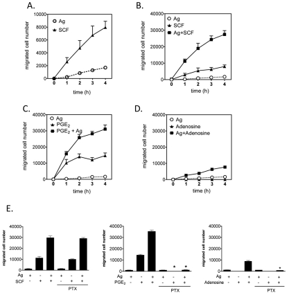
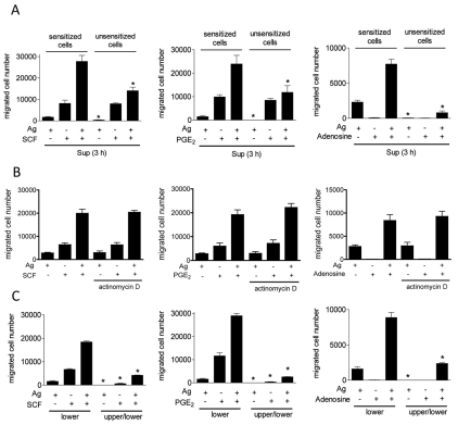
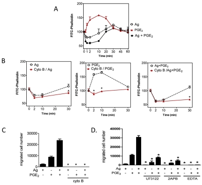
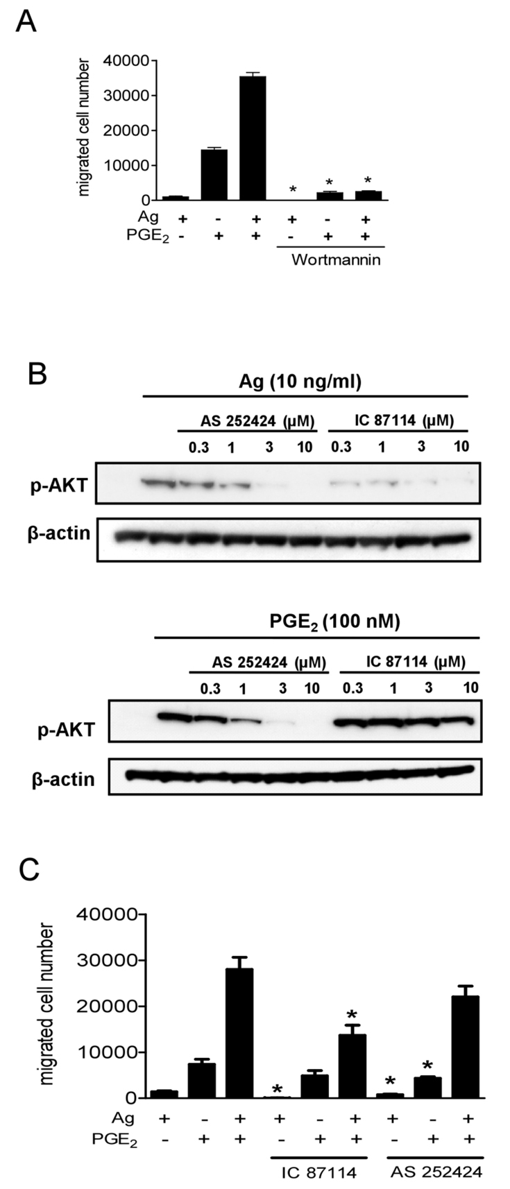
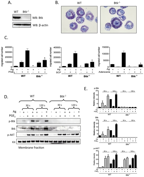
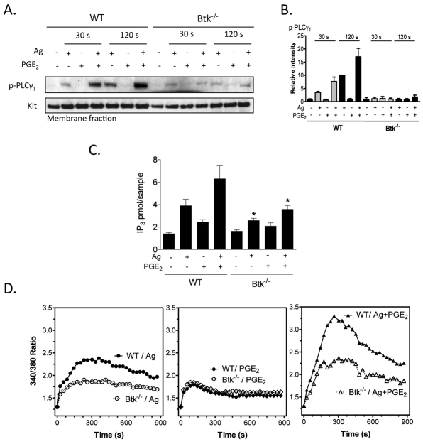
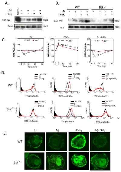
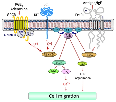
Mast cells infiltrate the sites of inflammation associated with chronic atopic disease and during helminth and bacterial infection. This process requires receptor-mediated cell chemotaxis across a concentration gradient of their chemotactic ligands. In vivo, mast cells are likely to be exposed to several such agents, which can cooperate in a synergistic manner to regulate mast cell homing. Here, we report that chemotaxis of mouse bone-marrow-derived mast cells (BMMCs) in response to the chemoattractants stem-cell factor (SCF) and prostaglandin (PG)E(2), is substantially enhanced following antigen-dependent ligation of the high-affinity receptor for IgE (FcεRI). These responses were associated with enhanced activation of phosphoinositide 3-kinase (PI3K), and downstream activation of the tyrosine protein kinase Btk, with subsequent enhanced phospholipase (PL)Cγ-mediated Ca(2+) mobilization, Rac activation and F-actin rearrangement. Antigen-induced chemotaxis, and the ability of antigen to amplify responses mediated by SCF, adenosine and PGE(2) were suppressed following inhibition of PI3K, and were impaired in BMMCs derived from Btk(-/-) mice. There were corresponding decreases in the PLCγ-mediated Ca(2+) signal, Rac activation and F-actin rearrangement, which, as they are essential for BMMC chemotaxis, accounts for the impaired migration of Btk-deficient cells. Taken together, these data demonstrate that, by regulating signaling pathways that control F-actin rearrangement, Btk is crucial for the ability of antigen to amplify mast-cell chemotactic responses.


Figures

Fig. 1.
Fig. 1.
FcεRI aggregation synergistically enhances chemotactic responses elicited by KIT and GPCRs. (A-D) BMMCs were sensitized overnight with IgE in cytokine-free medium. In these, and other, studies described, 3×10⁵ cells were added to the upper chambers. The chemotaxis assay was performed as described in the Materials and Methods. (E) BMMCs were preincubated with pertussis toxin (1 μg/ml) for 4 hours then washed and used for the assay. The indicated agonists [used at optimal concentrations (see supplementary material Fig. S1), antigen, Ag (10 ng/ml), SCF (10 ng/ml), adenosine (1 μM), PGE2 (100 nM)] were added in the lower chamber, and the migrated BMMCs were counted after 4 hours. Results are means ± s.e. of three separate experiments. *P<0.05 for comparison with same stimulation in control group by Student's t-test.
Fig. 2.
Fig. 2.
Synergistic cell migration is primarily dependent on chemotaxis. (A) IgE-sensitized BMMCs were washed three times with HEPES buffer containing 0.5% BSA, and then cells were stimulated with indicated agonists [antigen, Ag (10 ng/ml), SCF (10 ng/ml), adenosine (1 μM), PGE2 (100 nM)]. After 3 hours, cell-free supernatants (Sup) from antigen and/or other agonist-stimulated BMMCs were applied to the lower chamber. Sensitized or unsensitized BMMCs were placed in the upper chambers. After incubation for 4 hours, BMMCs migrating to the lower chambers were collected and counted. (B) Sensitized BMMCs were preincubated with or without actinomycin D (5 μg/ml) for 30 minutes, and cell migration was measured. (C) To test whether the cell migration was chemotaxis or chemokinesis, indicated agonists were placed in the lower chamber or both upper and lower chambers. Sensitized BMMCs were placed in upper chambers. After 4 hours, BMMCs migrating to the lower chambers were collected and counted. Results are means ± s.e. of three separate experiments. *P<0.05 for comparison with the same stimulation in control group by Student's t-test.
Fig. 3.
Fig. 3.
The role of cytoskeletal reorganization and Ca2+ in mast-cell migration. (A,B) Sensitized BMMCs were preincubated with or without cytochalasin B (10 μM) for 20 minutes. After preincubation with cytochalasin B, cells were stimulated with indicated agonists [Ag (10 ng/ml), PGE2 (100 nM)] for the indicated time, fixed, then permeabilized. Actin rearrangement was measured by using FITC-labeled phalloidin staining of the cells, followed by flow cytometry. Data are mean values of fluorescence intensities of phalloidin staining. (C) Sensitized BMMCs were preincubated with or without Cytochalasin B (10 μM) for 20-30 minutes, and cell migration was measured. (D) Sensitized BMMCs were preincubated with U73122 (1 μM), 2APB (50 μM), or EDTA (5 mM) in upper chambers and placed in 600 μl HEPES buffer containing 0.5% BSA and indicated inhibitors for 30 minutes, and then upper chambers were placed in the lower chambers containing the indicated agonists. Results are means ± s.e. of three separate experiments. *P<0.05 for comparison with the same stimulation in control group by Student's t-test.
Fig. 4.
Fig. 4.
The role of PI3K in synergistic chemotactic responses. (A) Sensitized BMMCs were preincubated with or without wortmannin (100 nM), in the upper chamber and placed in 600 μl HEPES buffer containing 0.5% BSA and wortmannin for 30 minutes. The upper chambers were then replaced in the lower chambers containing the indicated agonists. (B) Sensitized BMMCs were preincubated with indicated inhibitors for 20 minutes and then stimulated with the indicated agonists for 5 minutes. Following electrophoresis and membrane transfer, proteins were probed using anti-phospho-AKT (Ser473-P). To normalize protein loading, membranes were stripped and probed for β-actin, or alternatively identically loaded samples were probed for β-actin. The data shown are from three separate experiments, each repeated at least three times, with identical results, on separate cell preparations. (C) Sensitized BMMCs were preincubated with or without AS 252424 (3 μM) or IC 87114 (3 μM) in the upper chambers and placed in 600 μl HEPES buffer containing 0.5% BSA and indicated inhibitors for 30 minutes, and then upper chambers were replaced in the lower chamber containing the indicated agonists. Results in A and C are means ± s.e. of three separate experiments. *P<0.05 for comparison with the same stimulation in control group by Student's t-test.
Fig. 5.
Fig. 5.
The role of Btk in synergistic chemotactic responses. (A) To confirm knock out of Btk, cell lysates were prepared from WT, Btk−/− BMMCs and proteins were probed using an anti-Btk antibody. (B) 4-week-old BMMCs from WT and Btk−/− mice were stained with Toluidine Blue and images were obtained with an original magnification of 100×. (C) Sensitized WT and Btk−/− BMMCs were washed and placed in the upper chambers. Indicated agonists [Ag (10 ng/ml), SCF (10 ng/ml), PGE2 (100 nM), adenosine (1 μM)] were added to the lower chamber. Migrated cells were counted after incubation for 4 hours. (D) Sensitized WT and Btk−/− BMMCs were stimulated with indicated agonists [antigen, Ag (10 ng/ml), PGE2 (100 nM)] for 30 seconds or 2 minutes, and then membrane fractions were prepared to analyse the activation status. Following electrophoresis and membrane transfer, proteins were probed using the following antibodies: anti-phospho-Btk (Tyr551-P), anti-Btk, anti-phosphorylated AKT (Ser473-P). To normalize protein loading, membranes were probed for KIT. (E) Data were generated by scanning the blots in three independent experiments, and then normalizing to the response at 2 minutes obtained with Ag in WT BMMCs. Results are means ± s.e. of three separate experiments. *P<0.05 for comparison with the same stimulation in WT BMMCs by Student's t-test.
Fig. 6.
Fig. 6.
The role of Btk in PGE2-enhanced, antigen-mediated Ca2+ signaling. (A) Sensitized WT and Btk−/− BMMCs were stimulated with the indicated agonists [antigen, Ag (10 ng/ml), PGE2 (100 nM)] for 30 seconds or 2 minutes, and then membrane fractions were prepared. Following electrophoresis and membrane transfer, proteins were probed using anti-phospho-PLCγ (Tyr783-P). To normalize protein loading, membranes were probed for KIT. (B) The data were generated by scanning the blots from three independent experiments, and then normalizing to the response at 2 minutes obtained with Ag in WT BMMCs. The data are presented as the means + s.e. of three separate experiments. (C) WT and Btk−/− BMMCs were stimulated with antigen (Ag, 10 ng/ml), PGE2 (100 nM) or Ag and PGE2. After 30 seconds, the samples were processed and the Ins(1,4,5)P3 levels determined. The data are presented as means + s.e. of four separate experiments conducted in duplicate. (D) WT Btk−/− BMMCs were loaded with Fura-2 AM, and then changes in intracellular Ca2+ levels were determined after challenge with Ag (10 ng/ml) or PGE2 (100 nM) or Ag and PGE2. The Ca2+ data are from three representative experiments conducted on separate cell preparations. Results are means ± s.e. of three separate experiments. *P<0.05 for comparison with same stimulation in WT by Student's t-test.
Fig. 7.
Fig. 7.
The role of Btk in PGE2-enhanced, antigen-mediated cytoskeletal reorganization. (A,B) Rac activation was determined using a PAK-1 PBD assay kit. BMMCs were stimulated with indicated agonists [antigen, Ag (10 ng/ml) or PGE2 (100 nM)] for 2 minutes and lysates were mixed with GST-PBD bound to glutathione-agarose, and incubated for 4 hours at 4°C. For a positive control, cell lysates were incubated with 100 μM GTPγS. Precipitates were washed and suspended in sample buffer. Proteins were separated and blotted with an anti-Rac1 antibody. To establish equal Rac content in the reactions, cell lysates from each sample were probed by immunoblot analysis prior to reaction with GST-PBD. The western blots represent data from four separate experiments. (C-E) Cells were stimulated with indicated agonist Ag (10 ng/ml), PGE2 (100 nM) for indicated time (C) or 2 minutes (D,E), and then fixed and permeabilized. Actin polymerization was measured using FITC-labeled phalloidin staining of the cells, followed by flow cytometry (C,D) and imaging (E). Data are from four representative experiments conducted on separate cell preparations.
Fig. 8.
Fig. 8.
Proposed integrated signaling pathway responsible for the synergistic chemotactic responses elicited by antigen in the presence of SCF or GPCR agonists. For clarity, the pathways by which GPCRs and KIT regulate chemotaxis in the absence of FcεRI aggregation have not been included. Dotted lines indicate an amplification pathway and does not imply direct regulation. Concurrent ligation of KIT or GPCRs with FcεRI aggregation leads to enhanced activation of PI3Ks, which would result in elevated production of phosphoinositide-3,4,5-trisphosphate [PtdIns(3,4,5)P3 or PIP3] from phosphoinositide-4,5-bisphosphate [PtdIns(4,5)P2 or PIP2]. This would allow the synergistic translocation and activation of Btk (Fig. 5D). This in turn induces an enhancement in the Ca2+ signal and Rac-dependent F-actin rearrangement, which in combination lead to the observed synergy in mast-cell chemotaxis.

Similar articles

Cited by

References

    1. Ali H., Cunha-Melo J. R., Saul W. F., Beaven M. A. (1990). Activation of phospholipase C via adenosine receptors provides synergistic signals for secretion in antigen-stimulated RBL-2H3 cells. Evidence for a novel adenosine receptor. J. Biol. Chem. 265, 745-753 - PubMed
    1. Ali K., Camps M., Pearce W. P., Ji H., Ruckle T., Kuehn N., Pasquali C., Chabert C., Rommel C., Vanhaesebroeck B. (2008). Isoform-specific functions of phosphoinositide 3-kinases: p110 delta but not p110 gamma promotes optimal allergic responses in vivo. J. Immunol. 180, 2538-2544 - PMC - PubMed
    1. Allen J. D., Jaffer Z. M., Park S. J., Burgin S., Hofmann C., Sells M. A., Chen S., Derr-Yellin E., Michels E. G., McDaniel A., et al. (2009). p21-activated kinase regulates mast cell degranulation via effects on calcium mobilization and cytoskeletal dynamics. Blood 113, 2695-2705 - PMC - PubMed
    1. Brown J. M., Wilson T. M., Metcalfe D. D. (2008). The mast cell and allergic diseases: role in pathogenesis and implications for therapy. Clin. Exp. Allergy 38, 4-18 - PubMed
    1. de Gorter D. J., Beuling E. A., Kersseboom R., Middendorp S., van Gils J. M., Hendriks R. W., Pals S. T., Spaargaren M. (2007). Bruton's tyrosine kinase and phospholipase Cgamma2 mediate chemokine-controlled B cell migration and homing. Immunity 26, 93-104 - PubMed

Publication types

MeSH terms