

Prevalence of ponticulus posticus in Indian orthodontic patients
- PMID: 20587651
- PMCID: PMC3520248
- DOI: 10.1259/dmfr/16271087
Prevalence of ponticulus posticus in Indian orthodontic patients
Abstract
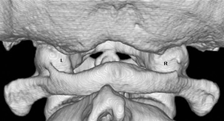
Objectives: The purpose of this study was to investigate the prevalence of complete ponticulus posticus in Indian orthodontic patients.
Methods: The presence and types of ponticuli posticus were investigated on 858 lateral cephalograms.
Results: Complete ponticulus posticus was found in 4.3% of the subjects studied with a male (5.33%) predominance over female in the population (3.76%).
Conclusions: Ponticulus posticus is not a rare anomaly and the patient must be told of the implications and importance of detecting ponticulus posticus on a lateral cephalogram. This information can prove beneficial for the diagnosis of head and neck symptoms later.
Figures


References
-
- Hassel B, Farman AG. Skeletal maturation evaluation using cervical vertebrae. Am J Orthod Dentofac Orthop 1995;107:58–66 - PubMed
-
- Baccetti T, Franchi L, McNamara JA., Jr The cervical vertebrae maturation (CVM) method for assessment of optimal treatment timing in dentofacial orthopedics. Sem Orthod 2005;11:119–129
-
- Soni P, Sharma V, Sengupta J. Cervical vertebral anomalies: incidental findings on lateral cephalograms. Angle Orthod 2008;78:176–180 - PubMed
-
- Farman AG, Escobar V. Radiographic appearance of the cervical vertebrae in normal and abnormal development. Br J Oral Surg 1982;20:264–74 - PubMed
-
- Ghanayem AJ, Paxinos O. Functional anatomy of joints, ligaments and disc. Clark CR (ed. In: ) The cervical spine (4th edn). Philadelphia Lippincotth Williams and Wilkins, 2005, pp 46–54.
