

Unusual radiographic appearance of ossifying fibroma in the left mandibular angle
- PMID: 20587656
- PMCID: PMC3520244
- DOI: 10.1259/dmfr/81820042
Unusual radiographic appearance of ossifying fibroma in the left mandibular angle
Abstract
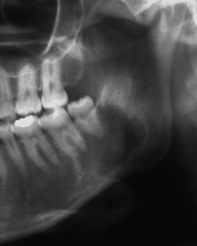
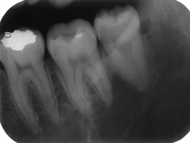
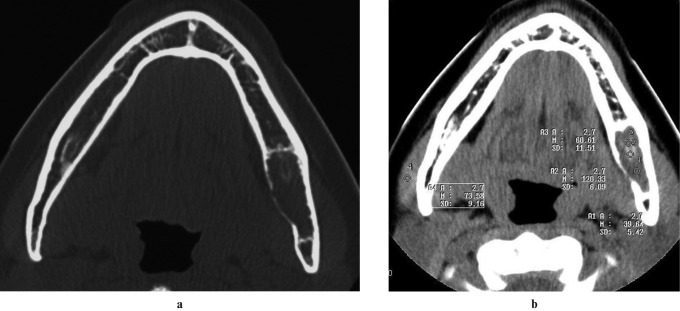
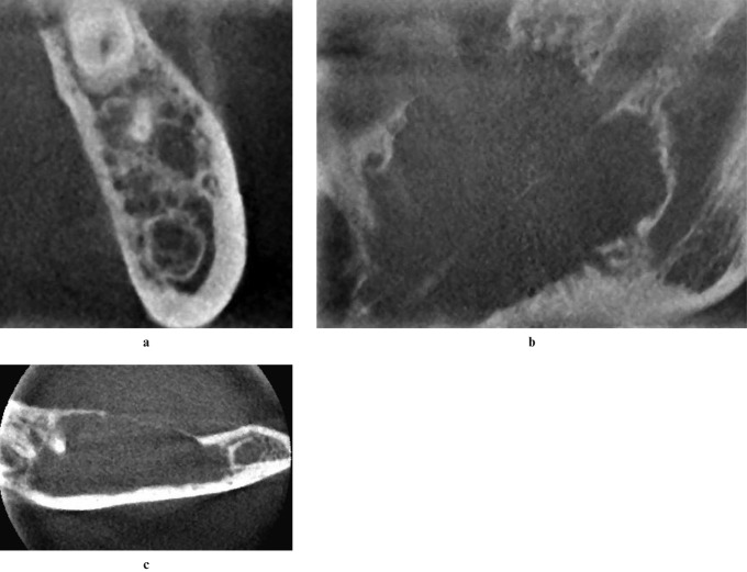
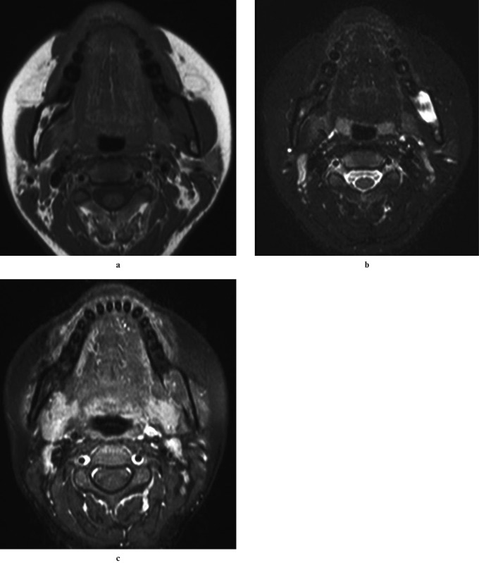
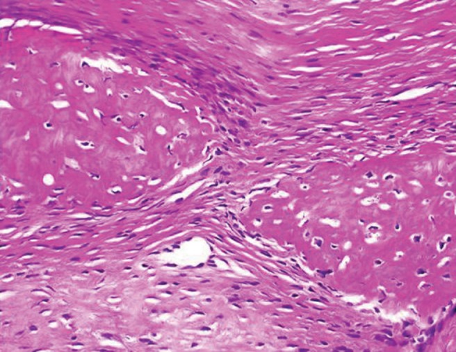
Ossifying fibroma is usually a unilocular lesion with a well-defined, thinly corticated margin radiographically, although various patterns have been noted. The patient was a 27-year-old woman with a painless radiolucent lesion demonstrated on panoramic radiography to involve the root-apex area of the left lower second and third molars. Radiographically, the lesion had some features of a benign tumour, such as an odontogenic myxoma. However, the deep invaginations towards the interalveolar septa suggested a simple bone cyst, whereas the irregular margin and lack of expansion or mandibular canal displacement were consistent with a malignant lesion. A hard tissue component was confirmed only by soft-tissue mode CT. Although this lesion was histopathologically diagnosed as ossifying fibroma, the conflicting imaging findings were challenging and very intriguing.
Figures
Similar articles
-
Central odontogenic fibroma with osteoid formation showing atypical radiographic appearance.Dentomaxillofac Radiol. 2009 Sep;38(6):426-30. doi: 10.1259/dmfr/28183053. Dentomaxillofac Radiol. 2009. PMID: 19700538
-
An ossifying fibroma presenting as Stafne's idiopathic bone cavity.Dentomaxillofac Radiol. 1997 Nov;26(6):361-3. doi: 10.1038/sj.dmfr.4600294. Dentomaxillofac Radiol. 1997. PMID: 9482014 Review.
-
Cemento-ossifying fibroma of the mandible.Dentomaxillofac Radiol. 1997 Jul;26(4):246-8. doi: 10.1038/sj.dmfr.4600245. Dentomaxillofac Radiol. 1997. PMID: 9442617
-
Distinguishing features of focal cemento-osseous dysplasia and cemento-ossifying fibromas. II. A clinical and radiologic spectrum of 316 cases.Oral Surg Oral Med Oral Pathol Oral Radiol Endod. 1997 Nov;84(5):540-9. doi: 10.1016/s1079-2104(97)90271-7. Oral Surg Oral Med Oral Pathol Oral Radiol Endod. 1997. PMID: 9394387
-
Odontogenic myxoma with diffuse calcifications: a case report and review of a rare histologic feature.Oral Surg Oral Med Oral Pathol Oral Radiol. 2016 Oct;122(4):e116-24. doi: 10.1016/j.oooo.2015.12.009. Epub 2016 Jan 3. Oral Surg Oral Med Oral Pathol Oral Radiol. 2016. PMID: 26948020 Review.
Cited by
-
Cone-beam computed tomography in the management of dentigerous cyst of the jaws: A report of two cases.Indian J Radiol Imaging. 2013 Oct;23(4):342-6. doi: 10.4103/0971-3026.125614. Indian J Radiol Imaging. 2013. PMID: 24604939 Free PMC article.
-
Imaging in the diagnosis of cemento-ossifying fibroma: a case series.J Clin Imaging Sci. 2012;2:52. doi: 10.4103/2156-7514.100373. Epub 2012 Aug 30. J Clin Imaging Sci. 2012. PMID: 23029635 Free PMC article.
-
Ossifying fibroma in the mandibular angle mimicking metastatic clear cell renal cell carcinoma: A case report.Medicine (Baltimore). 2019 Aug;98(33):e16595. doi: 10.1097/MD.0000000000016595. Medicine (Baltimore). 2019. PMID: 31415350 Free PMC article.
References
-
- Summerlin DJ, Tomich CE. Focal cemento-osseous dysplasia: a clinicopathologic study of 221 cases. Oral Surg Oral Med Oral Pathol 1994;78:611–620 - PubMed
-
- Voytek TM, Ro JY, Edeiken J, Ayala AG. Fibrous dysplasia and cemento-ossifying fibroma. A histologic spectrum. Am J Surg Pathol 1995;19:775–781 - PubMed
-
- Su L, Weathers DR, Waldron CA. Distinguishing features of focal cemento-osseous dysplasia and cemento-ossifying fibromas. II. A clinical and radiologic spectrum of 316 cases. Oral Surg Oral Med Oral Pathol 1997;84:540–548 - PubMed
-
- MacDonald-Jankowski DS. Focal cemento-osseous dysplasia: a systematic review. Dentomaxillofac Radiol 2008;37:350–360 - PubMed
-
- Langlais RP, Langland OE, Nortje CH. Diagnostic imaging of the jaws. Baltimore: Williams & Wilkins, 1995
Publication types
MeSH terms
Substances
LinkOut - more resources
Full Text Sources
Miscellaneous
