. 2010 Nov;47(6):1028-39.
doi: 10.1177/0300985810374842. Epub 2010 Jun 29.

The haploinsufficient Col3a1 mouse as a model for vascular Ehlers-Danlos syndrome

Affiliations

The haploinsufficient Col3a1 mouse as a model for vascular Ehlers-Danlos syndrome

T K Cooper et al. Vet Pathol. 2010 Nov.

Abstract

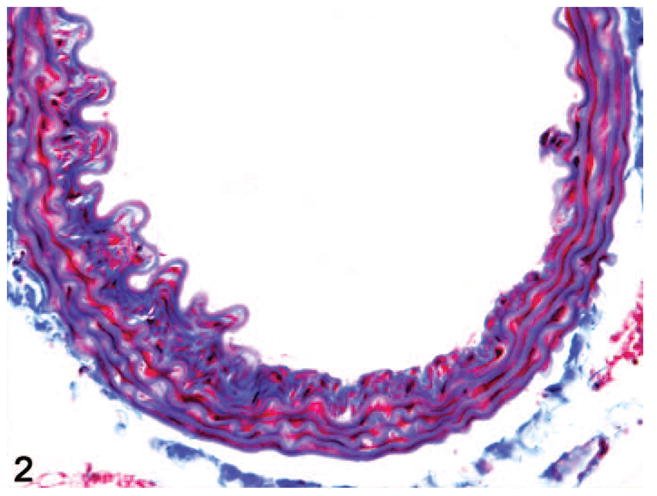
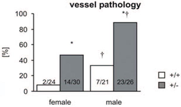
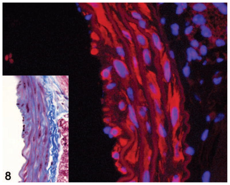
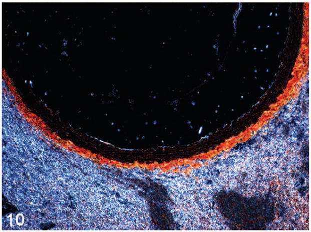
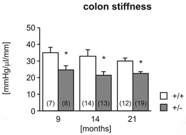
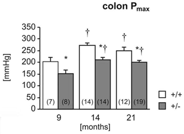
Vascular Ehlers-Danlos syndrome is a rare genetic disorder resulting from mutations in the α-1 chain of type III collagen (COL3A1) and manifesting as tissue fragility with spontaneous rupture of the bowel, gravid uterus, or large or medium arteries. The heterozygous Col3a1 knockout mouse was investigated as a model for this disease. The collagen content in the abdominal aorta of heterozygotes was reduced, and functional testing revealed diminishing wall strength of the aorta in these mice. Colons were grossly and histologically normal, but reduced strength and increased compliance of the wall were found in heterozygotes via pressure testing. Although mice demonstrated no life-threatening clinical signs or gross lesions of vascular subtype Ehlers-Danlos syndrome type IV, thorough histological examination of the aorta of heterozygous mice revealed the presence of a spectrum of lesions similar to those observed in human patients. Lesions increased in number and severity with age (0/5 [0%] in 2-month-old males vs 9/9 [100%] in 14-month-old males, P < .05) and were more common in male than female mice (23/26 [88.5%] vs 14/30 [46.7%] in 9- to 21-month-old animals, P < .05). Haploinsufficiency for Col3a1 in mice recapitulates features of vascular Ehlers-Danlos syndrome in humans and can be used as an experimental model.


Conflict of interest statement

Declaration of Conflict of Interest

The authors declared that they had no conflicts of interests with respect to their authorship or the publication of this article.

Figures

Figure 1
Figure 1
Aorta. Grade 2 lesion with a large defect in the internal elastic lamina (IEL) and significant subintimal spindle cell proliferation with deposition of collagen. Masson’s trichrome.
Figure 2
Figure 2
Aorta. Grade 3 lesion with a larger IEL defect, more florid spindle cell proliferation, and extensive deposition of collagenous matrix. Masson’s trichrome.
Figure 3
Figure 3
Aorta. Grade 4 lesion with fragmentation of IEL as well as multiple medial elastic laminae, extensive medial interlamellar deposition of collagen, and jumbled irregular elastic fibers at the edge of a focally extensive attenuation in the thickness of the aortic wall. Masson’s trichrome.
Figure 4
Figure 4
Aorta. A thick eccentric subintimal plaque of spindle cells and collagenous matrix segmentally expands the inner aortic wall and narrows the lumen. Outer media and adventitia are unremarkable. Masson’s trichrome.
Figure 5
Figure 5
Comparison of the presence or absence of aortic pathology (lesions > grade 2) in 9- to 21-month-old animals demonstrates a significant association with genotype and sex (*P < .05 vs wild-type, †P < .05 vs female).
Figure 6
Figure 6
Evaluation of the presence or absence of aortic pathology (lesions > grade 2) in each age group demonstrates a significant association with age, sex, and genotype (*P < .05 vs wild-type, †P < .05 vs female).
Figure 7
Figure 7
Cumulative lesion score (lesions > grade 2) in each age group demonstrates a significant association with age, sex, and genotype (*P < .05 vs all female or male wild-type (respectively), †P < .05 vs female, #P < .05 vs 5 months).
Figure 8
Figure 8
Aorta. Smoothelin immunofluorescence of a grade 2 lesion showing smoothelin-positive (red staining) and smoothelin-negative spindle cells within the media. Alexa Fluor 594 conjugate with DAPI counterstain. Inset shows histology of the same lesion. Masson’s trichrome.
Figure 9
Figure 9
Abdominal aorta of wild-type (Fig. 9) and heterozygote (Fig. 10) mice stained with picro-sirius red and viewed under polarized light demonstrates reduced adventitial and, to a lesser extent medial, total collagen in the heterozygote, consistent with genotype.
Figure 10
Figure 10
Abdominal aorta of wild-type (Fig. 9) and heterozygote (Fig. 10) mice stained with picro-sirius red and viewed under polarized light demonstrates reduced adventitial and, to a lesser extent medial, total collagen in the heterozygote, consistent with genotype.
Figure 11
Figure 11
By picro-sirius red staining, wild-type (WT) mice had significantly higher collagen per unit area than heterozygotes (HT) in the abdominal aorta. AC, ascending aorta; Trn, transverse aorta; Tho, thoracic aorta; Abd, abdominal aorta (*P < .05 vs WT).
Figure 12
Figure 12
Western blotting of isolated abdominal aorta shows significant reduction of type III collagen levels in HT compared with age- and sex-matched WT littermates.
Figure 13
Figure 13
Evaluation of type I collagen carboxy-terminal peptide from 24-hour urine (normalized to creatinine) in 9-month-old animals demonstrates significantly higher urinary PICP in male HT mice relative to WT males and to females (P < .05). There is no significant difference between WT and HT females.
Figure 14
Figure 14
The maximum pressure in the abdominal aorta was measured in 14- and 21-month-old wild-type (+/+) and heterozygous (+/−) mice. Number of experiments shown in parentheses.
Figure 15
Figure 15
The compliance of arteria gracilis was measured using isobaric myography in 10-month-old wild-type (+/+) and heterozygous (+/−) mice (n = 7). *P <.05 vs wild-type.
Figure 16
Figure 16
Biomechanical properties of the colon. Colon stiffness (Fig. 16) and maximal holding pressure (Fig. 17) were measured in 9-, 14-, and 21-month-old wild-type (+/+) and heterozygous (+/−) mice. Number of experiments shown in parentheses. *P < .05 vs wild-type, †P <.05 vs 9 months.
Figure 17
Figure 17
Biomechanical properties of the colon. Colon stiffness (Fig. 16) and maximal holding pressure (Fig. 17) were measured in 9-, 14-, and 21-month-old wild-type (+/+) and heterozygous (+/−) mice. Number of experiments shown in parentheses. *P < .05 vs wild-type, †P <.05 vs 9 months.

References

    1. Arteaga-Solis E, Gayraud B, Ramirez F. Elastic and collagenous networks in vascular diseases. Cell Struct Funct. 2000;25:69–72. - PMC - PubMed
    1. Barabas AP. Ehlers-Danlos syndrome type IV. N Engl J Med. 2000;343:366. author reply 368. - PubMed
    1. Baxter BT. Heritable diseases of the blood vessels. Cardiovasc Pathol. 2005;14:185–188. - PubMed
    1. Beighton P, De Paepe A, Steinmann B, Tsipouras P, Wenstrup RJ. Ehlers-Danlos syndromes: revised nosology, Villefranche, 1997. Ehlers-Danlos National Foundation (USA) and Ehlers-Danlos Support Group (UK) Am J Med Genet. 1998;77:31–37. - PubMed
    1. Blaker H, Funke B, Hausser I, Hackert T, Schirmacher P, Autschbach F. Pathology of the large intestine in patients with vascular type Ehlers-Danlos syndrome. Virchows Arch. 2007;450:713–717. - PubMed

Publication types

MeSH terms

Substances