. 2010 Sep 1;21(17):3007-16.
doi: 10.1091/mbc.E10-03-0232. Epub 2010 Jun 29.

The Rsr1/Bud1 GTPase interacts with itself and the Cdc42 GTPase during bud-site selection and polarity establishment in budding yeast

Affiliations

The Rsr1/Bud1 GTPase interacts with itself and the Cdc42 GTPase during bud-site selection and polarity establishment in budding yeast

Pil Jung Kang et al. Mol Biol Cell. .

Abstract

Cell polarization occurs along a single axis that is generally determined in response to spatial cues. In budding yeast, the Rsr1 GTPase and its regulators direct the establishment of cell polarity at the proper cortical location in response to cell type-specific cues. Here we use a combination of in vivo and in vitro approaches to understand how Rsr1 polarization is established. We find that Rsr1 associates with itself in a spatially and temporally controlled manner. The homotypic interaction and localization of Rsr1 to the mother-bud neck and to the subsequent division site are dependent on its GDP-GTP exchange factor Bud5. Analyses of rsr1 mutants suggest that Bud5 recruits Rsr1 to these sites and promotes the homodimer formation. Rsr1 also exhibits heterotypic interaction with the Cdc42 GTPase in vivo. We show that the polybasic region of Rsr1 is necessary for the efficient homotypic and heterotypic interactions, selection of a proper growth site, and polarity establishment. Our findings thus suggest that dimerization of GTPases may be an efficient mechanism to set up cellular asymmetry.


Figures
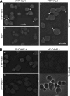
Figure 1.
Figure 1.
Rsr1 associates with itself and Cdc42 in vivo. (A) BiFC assays in haploid α cells (HPY1200), which express YFPN-Rsr1 from the RSR1 locus and carry YCpYFPC-RSR1 (a) or YCpYFPC-rsr1Δ (c); and in diploid a/α cells coexpressing YFPN-Rsr1 and YFPC-Rsr1 (HPY1214; b). Arrowheads mark cells with clear BiFC signals. Pixel intensity of the cell marked with an arrow is shown in comparison to other cells in Figure 5D. Images were captured with the YFP filter for 6-s exposure and deconvolved. Bar, 5 μm. (B) BiFC assays in diploid a/α cells coexpressing YFPN-Rsr1 and VC-Cdc42 (HPY1209) (a); VN-Cdc42 and VC-Cdc42 (HPY1198) (b); and YFPN-Rsr1KS and VC-Cdc42 (HPY1217) (c). Phase images are also shown below for the same cells of HPY1198. Images were captured with the YFP filter for 8-s exposure and deconvolved. Bar, 5 μm.
Figure 2.
Figure 2.
Bud5 is necessary for efficient homotypic interaction of Rsr1. BiFC assays were done in a/α wild-type cells coexpressing YFPN-Rsr1G12V and YFPC-Rsr1G12V (HPY1568; a); and YFPN-Rsr1K16N and YFPC-Rsr1K16N (HPY1608; b). Similarly, BiFC assays were done in abud5Δ/bud5Δ cells coexpressing YFPN-Rsr1 and YFPC-Rsr1 (HPY1575; c); and YFPN-Rsr1K16N and YFPC-Rsr1K16N (HPY1824; d). Images were captured and processed as in Figure 1A. Bars, 5 μm.
Figure 3.
Figure 3.
Localization of YFP-Rsr1 to the mother-bud neck occurs after anaphase and is dependent on Bud5. Localization of YFP-Rsr1 and Tub1-CFP in haploid wild-type (HPY1632), bud5Δ (HPY1668), and bud2Δ (HPY1664) cells. YFP-Rsr1 and Tub1-CFP were expressed from the RSR1 and URA3 loci on the chromosomes, respectively. A cell marked with an arrow (a) undergoes nuclear division; and cells marked with arrowheads (b–d) have completed nuclear division. Pixel intensities of these cells are shown in Supplemental Figure S1. A series of Z-sections were captured with the YFP and CFP filters for YFP-Rsr1 and Tub1-CFP, respectively. Images were deconvolved, and a single Z section is shown. Bar, 5 μm.
Figure 4.
Figure 4.
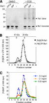
Rsr1 forms a dimer in vitro. (A) Purified Rsr1 (10 μg/ml) was preloaded with GTPγS or GDP, or in nucleotide-empty state (−), recovered after cleaving off the GST moiety, treated with 0.1 mM EGS or mock-treated (DMSO), and then subjected to SDS-PAGE. Rsr1 was detected with polyclonal antibodies against Rsr1. (B) Rsr1 forms a stable homodimer in solution. Purified Rsr1 (50 μl; 0.05 mg/ml) was applied to a size exclusion chromatography column (Sephacryl-200HR; 1.6 ml bed volume) after preloading with [3H]GDP (•) or [3H]GTP (▵). The molecular-weight standards, BSA (67 kDa) and GST (26 kDa), eluted in fractions 13–14 and 26–27, respectively. Fractions 14–16 and 24–25 contain most of the Rsr1 dimers and monomers, respectively. The amount of [3H]GDP or [3H]GTP bound to Rsr1 was measured by scintillation counting (dpm), and plotted after subtracting the background dpm. (C) Dimerization of Rsr1 is concentration-dependent. Rsr1 (5 μl of each indicated concentration) preloaded with [3H]GDP was applied to a size exclusion chromatography column as in Figure 4B. An equal molar concentration of [3H]GDP was used for GDP loading of Rsr1, which was kept at each concentration.
Figure 5.
Figure 5.
Homotypic interaction of Rsr1 requires an intact polybasic region near the C terminus. (A) Rsr1K260–264S (Rsr1KS) binds mantGTP as efficiently as wild type. Purified GST-Rsr1 (∼2 μM; green line) and GST-Rsr1KS (∼3 μM; blue line) were used to test mantGTP binding. The amount of wild-type and Rsr1KS mutant proteins used for mantGTP binding was estimated by Coomassie blue staining (as shown in inset) with the BSA standards. Rsr1KS runs slightly faster than wild type in a protein gel, and a slower migrating band presumably corresponds to nascent protein with unmodified CaaX box. GST (∼3 μM; orange line) was used to detect background fluorescence. MantGTP was excited at 360 nm, and emission spectra were collected from 400 to 600 nm. (B) HA-Rsr1 interacts with GST-Rsr1 but not with GST-Rsr1KS in vitro. HA-Rsr1, immunoprecipitated from yeast extract, was incubated with purified GST-Rsr1 or GST-Rsr1KS, preloaded with GTPγS or GDP. GST-Rsr1 or GST-Rsr1KS was detected with polyclonal antibodies against GST (top panel); and HA-Rsr1 was detected with a mAb against HA epitope (bottom panel). Purified GST-Rsr1 and GST-Rsr1KS added in the binding reaction are shown (input). (C) BiFC assays were performed in diploid a/α cells coexpressing YFPN-Rsr1KS and YFPC-Rsr1KS (HPY1220) grown at 25°C (a) or 30°C (b); and YFPN-Rsr1 and YFPC-Rsr1KS (HPY1219) at 25°C (c) or 30°C (d). Images were captured and processed as in Figure 1A. Size bar, 5 μm. Note: Although cells were grown at the indicated temperatures, these cells were kept at the same, room temperature during microscopic observation. This might have contributed to some faint BiFC signal observed in the cells grown at 30°C (b). (D) Pixel intensity is shown along the line for the cells marked with arrows in Figure 1Ab and Figure 5C, a and b.
Figure 6.
Figure 6.
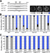
The intact PBR of Rsr1 is necessary for bud-site selection. (A) The carboxy-terminal region of Rsr1 and the mutations in the PBR are shown. Each rsr1 mutant carries substitutions of Lys to Ser (underlined). (B) Budding patterns of haploid wild-type (HPY11), rsr1Δ (HPY263), rsr1-7 (HPY1625), rsr1-8 (HPY1741), and rsr1-9 (HPY1740) cells, grown at the indicated temperature, were determined after Calcofluor staining. Each rsr1 mutant was expressed from its native promoter at the RSR1 locus. About 300–400 cells were counted for each strain in three independent sets of experiments. The average percentage of each budding pattern is shown (SD <2%); axial (gray bar), bipolar (black bar), and random (blue bar). Calcofluor staining of a representative cell budding in each pattern is shown above the graph. (C) Budding pattern of wild-type cells (HPY11) carrying each RSR1 plasmid (on YEplac195) or empty vector was determined as in Figure 6B. RSR1G12V, KS and RSR1K16N, KS represent the RSR1 plasmids carrying the K260-264S mutation (rsr1-7KS) as well as each dominant mutation. Three independent transformants of each plasmid were grown overnight in SD-URA media at 30 or 37°C, and their budding patterns were determined after Calcofluor staining. About 300 cells of each sample were counted twice and the mean (%) is shown; axial (gray bar), bipolar (black bar), and random (blue bar) pattern.
Figure 7.
Figure 7.
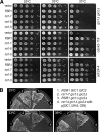
The rsr1-7KS mutant is defective in polarity establishment. (A) The gic1Δ gic2Δ mutant (HPY1523) (top panel) and the cdc42-118 mutant (DDY1326) (middle panel) carrying each RSR1 plasmid (on YEplac195) or vector control were grown at 25, 33, and 37°C for 3 d on SC-URA. Similarly, the cdc24-4 mutant (Y147) carrying the same set of the RSR1 plasmids was plated on SC-URA containing 1 M sorbitol (bottom panel). Each spot of cells represented a 10-fold serial dilution from left to right (starting from OD600 = 0.5). (B) The rsr1-7 allele expressed from the RSR1 locus can rescue the lethality of rsr1Δ gic1Δ gic2Δ at 25 but not at 36°C. All strains are isogenic to HPY210: (1) wild type (HPY210); (2) rsr1-7 gic1Δ gic2Δ (HPY1609); (3) RSR1 gic1Δ gic2Δ (YKT342); and (4) rsr1Δ gic1Δ gic2Δ (YKT401) carrying pKT1276 [GIC1, URA3, CEN] plasmid. Each strain was streaked on SC plates containing 5-FOA and incubated at 25, 33, and 36°C for 3 d.

Similar articles

Cited by

References

    1. Ausubel F. M., Brent R., Kingston R. E., Moore D. D., Seidman J. G., Struhl K. New York: John Wiley & Sons; 1999. Current Protocols in Molecular Biology.
    1. Bailly E., Cabantous S., Sondaz D., Bernadac A., Simon M.-N. Differential cellular localization among mitotic cyclins from Saccharomyces cerevisiae: a new role for the axial budding protein Bud3 in targeting Clb2 to the mother-bud neck. J. Cell Sci. 2003;116:4119–4130. - PubMed
    1. Beck R., Sun Z., Adolf F., Rutz C., Bassler J., Wild K., Sinning I., Hurt E., Brügger B., Béthune J., Wieland F. Membrane curvature induced by Arf1-GTP is essential for vesicle formation. Proc. Natl. Acad. Sci. USA. 2008;105:11731–11736. - PMC - PubMed
    1. Bender A. Genetic evidence for the roles of the bud-site-selection genes BUD5 and BUD2 in control of the Rsr1p (Bud1p) GTPase in yeast. Proc. Natl. Acad. Sci. USA. 1993;90:9926–9929. - PMC - PubMed
    1. Bender A., Pringle J. R. Multicopy suppression of the cdc24 budding defect in yeast by CDC42 and three newly identified genes including the ras-related gene RSR1. Proc. Natl. Acad. Sci. USA. 1989;86:9976–9980. - PMC - PubMed

Publication types

MeSH terms

Substances