. 2010 Aug 15;21(16):2832-43.
doi: 10.1091/mbc.E10-01-0059. Epub 2010 Jun 29.

Molecular characterization of EG-VEGF-mediated angiogenesis: differential effects on microvascular and macrovascular endothelial cells

Affiliations

Molecular characterization of EG-VEGF-mediated angiogenesis: differential effects on microvascular and macrovascular endothelial cells

Sophie Brouillet et al. Mol Biol Cell. .

Abstract

Endocrine gland derived vascular endothelial growth factor (EG-VEGF) also called prokineticin (PK1), has been identified and linked to several biological processes including angiogenesis. EG-VEGF is abundantly expressed in the highest vascularized organ, the human placenta. Here we characterized its angiogenic effect using different experimental procedures. Immunohistochemistry was used to localize EG-VEGF receptors (PROKR1 and PROKR2) in placental and umbilical cord tissue. Primary microvascular placental endothelial cell (HPEC) and umbilical vein-derived macrovascular EC (HUVEC) were used to assess its effects on proliferation, migration, cell survival, pseudovascular organization, spheroid sprouting, permeability and paracellular transport. siRNA and neutralizing antibody strategies were used to differentiate PROKR1- from PROKR2-mediated effects. Our results show that 1) HPEC and HUVEC express both types of receptors 2) EG-VEGF stimulates HPEC's proliferation, migration and survival, but increases only survival in HUVECs. and 3) EG-VEGF was more potent than VEGF in stimulating HPEC sprout formation, pseudovascular organization, and it significantly increases HPEC permeability and paracellular transport. More importantly, we demonstrated that PROKR1 mediates EG-VEGF angiogenic effects, whereas PROKR2 mediates cellular permeability. Altogether, these data characterized angiogenic processes mediated by EG-VEGF, depicted a new angiogenic factor in the placenta, and suggest a novel view of the regulation of angiogenesis in placental pathologies.


Figures

Figure 1.
Figure 1.
PROKR1 and PROKR2 protein expression in placental-tissue, umbilical cord and in isolated HPECs and HUVECs. (A) Chorionic villi and umbilical cord sections immunostained with anti-PROKR1 and anti-PROKR2 antibodies. The undersized photographs on the right show tissue sections incubated with the respective preimmune sera. Subset photographs in each panel represent higher magnifications for the staining in endothelial cells. CT, cytotrophoblast; Ho, Hofbauer cells; St, syncytiotrophoblast; Ec, endothelial cells; bv, blood vessels. Scale bar, 50 μm. (B) A representative Western blot analysis of PROKR1 and PROKR2 expression in HPECs and HUVECs. (C) A quantification of levels PROKR1 and PROKR2 protein expression in HPEC and HUVEC cells. *p < 0.05.
Figure 2.
Figure 2.
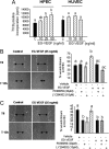
EG-VEGF increases HPEC but not HUVEC proliferation and migration. (A) [3H]Thymidine incorporation into HPEC and HUVEC cells, in the absence or presence of EG-VEGF. A significant increase of HPEC proliferation was observed with 25 and 50 ng/ml EG-VEGF (*p < 0.05). No significant effect was observed on HUVEC cells. (B and C) Photographs of wounded HPEC and HUVEC monolayers, respectively, at 0, and 12 h after wounding. The plots show percentages of wound closure after 12 h of treatment with EG-VEGF in the absence or presence of PD98059 and the LY294002, the inhibitors of MAP kinases and PI3K, respectively. Bars with different letters are significantly different from each other (p < 0.05).
Figure 3.
Figure 3.
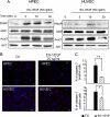
EG-VEGF is a survival factor for HPEC and HUVEC cells. (A) Representative Western blots of MAP kinase and AKT phosphorylations after treatment with EG-VEGF in HPEC and HUVEC cells. Standardization of the protein signals was done with antibodies against dephospho-MAP kinases and -AKT. (B) shows the effect of EG-VEGF on caspase 3 expression in HPEC and HUVEC cells after serum starvation and challenging with EG-VEGF (25 ng/ml). (C) The percentage of caspase 3–positive cells. Three randomly selected microscopic fields were observed, and ≥200 cells/field were evaluated. (**p < 0.01, *p < 0.05). Bar, 20 μm.
Figure 4.
Figure 4.
EG-VEGF increases HPEC but not HUVEC cord-like organization. (A) Photographs of HPEC and HUVEC cells cultured on Matrigel for 0 and 10 h in the absence or the presence of EG-VEGF (25 ng/ml). Note that EG-VEGF increased HPEC but not HUVEC organization into cord-like structures compared with the control condition. (B) Measurements of the number of branches formed by the cells after 10 h of culture in the absence or presence of EG-VEGF. *p < 0.05.
Figure 5.
Figure 5.
EG-VEGF, FGF, and VEGF effects on sprouting of HPEC spheroids. (A) Representative photographs of spheroids formed from HPEC cells and cultured in collagen gel for 0 or 12 h in the absence or presence of EG-VEGF (25 ng/ml), FGF-2 (25 ng/ml), and VEGF (100 ng/ml). Note that EG-VEGF increased HPEC spheroid sprouting compared with the control, FGF2, and VEGF conditions. (B) Quantification of the number of sprouts formed after 12 h in four independent experiments. (C) Representative photographs of spheroids formed from HUVEC cells and cultured in collagen gel for 0 and 12 h in the absence or the presence of EG-VEGF or VEGF (100 ng/ml). Note that EG-VEGF did not affect HUVEC spheroid sprouting compared with the control and VEGF conditions. Data represent the mean ± SEM (*p < 0.05, ***p < 0.001). Bar, 150 μm.
Figure 6.
Figure 6.
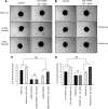
EG-VEGF angiogenic effects are mediated by PROKR1 and not PROKR2. (A and B) EG-VEGF (25 ng/ml) effect on spheroid sprouting of HPEC cells that had been silenced for PROKR1 and PROKR2 mRNA using siRNA (siRNA strategy), or treated with PROKR1 and R2 blocking antibodies (antibody strategy), respectively. (C and D) Quantifications of the number of sprouts in three independent experiments for both strategies. In the two sets of experiments, EG-VEGF significantly increased the number of sprouts. Both siRNA to PROKR1 and its blocking antibody inhibited EG-VEGF effect. However, nor siRNA to PROKR2, neither its blocking antibody did affect the spheroid sprouting. Data represent the mean ± SEM (***p < 0.001, **p < 0.01, ns, not significant). Bar, 100 μm.
Figure 7.
Figure 7.
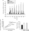
Effects of EG-VEGF, VEGF, and thrombin on the transendothelial electrical resistance (TEER) across HPEC monolayers. (A) The decrease in the TEER of HPEC cells after their incubation with EG-VEGF (25 ng/ml), VEGF (25 ng/ml), or thrombin (70 U/ml). Changes in resistance were measured at the time points 0, 5, 10, 15, 20, 25, and 35 min. Data represent the means ± SEM from three independent experiments. The results were normalized to the respective control. (*p < 0.05). (B). Effects of EG-VEGF and thrombin on the paracellular transport of [3H]mannitol in HPEC cells. The graph represents the plot of [3H]mannitol accumulation in the abluminal chamber of HPECs. (C) The permeability coefficient of EG-VEGF and thrombin that was calculated as described in Materials and Methods. Data represent the mean ± SEM from three independent experiments (*p < 0.05).
Figure 8.
Figure 8.
EG-VEGF effects on HPEC permeability are mediated by PROKR2 and not PROKR1: Panels (A) and (B) show EG-VEGF (25 ng/ml) effect on the permeability of HPEC cells that had been silenced for PROKR1 and PROKR2 mRNA using siRNA (siRNA strategy), or treated with PROKR1 and R2 blocking antibodies (antibody strategy), respectively. In the two sets of experiments, EG-VEGF significantly increased HPEC permeability. Both siRNA to PROKR2 and its blocking antibody inhibited EG-VEGF effect. However, nor siRNA to PROKR1, neither its blocking antibody did affect the permeability. Data represent the mean ± SEM. Bars with different letters are significantly different from each other (p < 0.05).

Similar articles

Cited by

References

    1. Burton G. J., Jauniaux E. Sonographic, stereological and Doppler flow velocimetric assessments of placental maturity. Br. J. Obstet. Gynaecol. 1995;102:818–825. - PubMed
    1. Charnock-Jones D. S., Burton G. J. Placental vascular morphogenesis. Baillieres Best Pract. Res. Clin. Obstet. Gynaecol. 2000;14:953–968. - PubMed
    1. Charnock-Jones D. S., Kaufmann P., Mayhew T. M. Aspects of human fetoplacental vasculogenesis and angiogenesis. I. Molecular regulation. Placenta. 2004;25:103–113. - PubMed
    1. Chen J., Kuei C., Sutton S., Wilson S., Yu J., Kamme F., Mazur C., Lovenberg T., Liu C. Identification and pharmacological characterization of prokineticin 2 beta as a selective ligand for prokineticin receptor 1. Mol. Pharmacol. 2005;67:2070–2076. - PubMed
    1. Dellian M., Witwer B. P., Salehi H. A., Yuan F., Jain R. K. Quantitation and physiological characterization of angiogenic vessels in mice: effect of basic fibroblast growth factor, vascular endothelial growth factor/vascular permeability factor, and host microenvironment. Am. J. Pathol. 1996;149:59–71. - PMC - PubMed

Publication types

MeSH terms

Substances

LinkOut - more resources