. 2010;2(6):560-75.
doi: 10.1159/000317134. Epub 2010 Jun 26.

Rapid neutrophil destruction following phagocytosis of Staphylococcus aureus

Affiliations

Rapid neutrophil destruction following phagocytosis of Staphylococcus aureus

Scott D Kobayashi et al. J Innate Immun. 2010.

Abstract

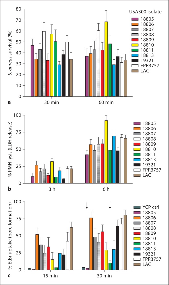
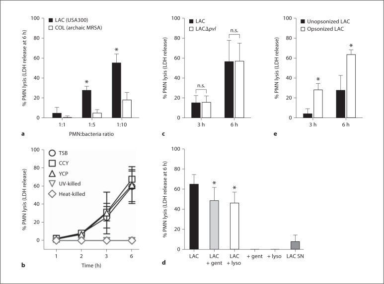
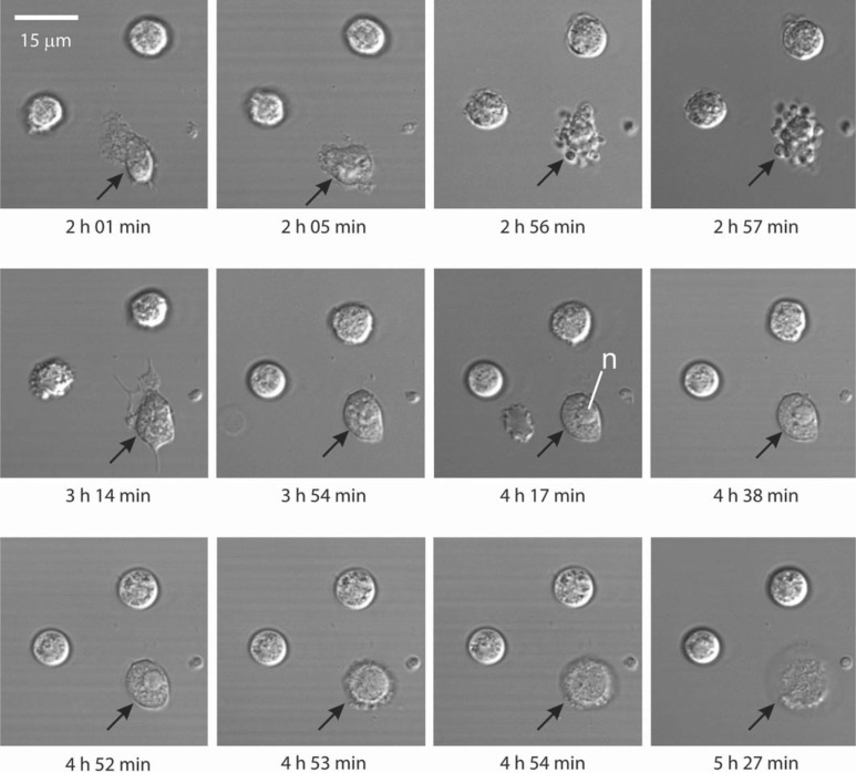
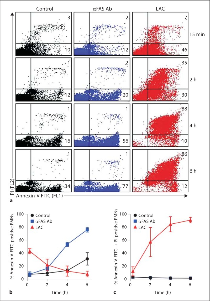
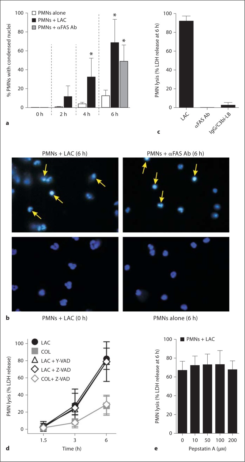
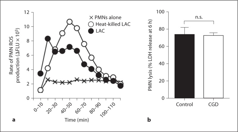
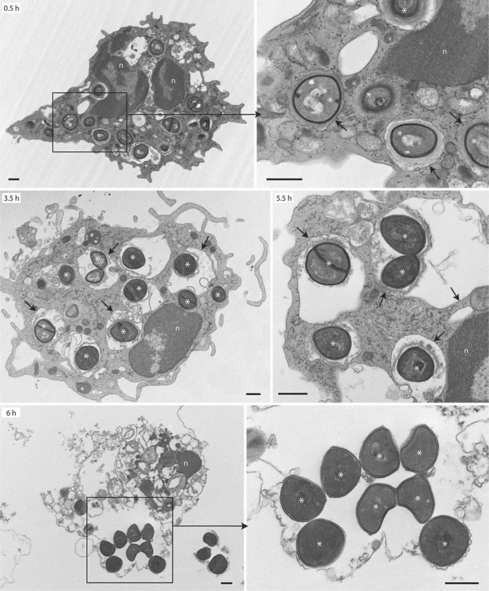
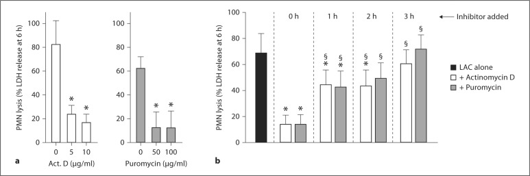
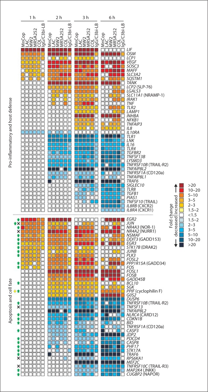
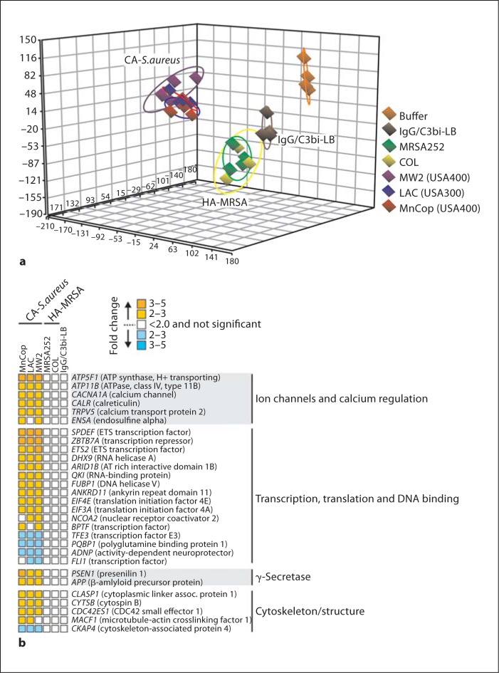
Mechanisms underlying the enhanced virulence phenotype of community-associated methicillin-resistant Staphylococcus aureus (CA-MRSA) are incompletely defined, but presumably include evasion of killing by human polymorphonuclear leukocytes (PMNs or neutrophils). To better understand this phenomenon, we investigated the basis of rapid PMN lysis after phagocytosis of USA300, a prominent CA-MRSA strain. Survival of USA300 clinical isolates after phagocytosis ultimately resulted in neutrophil lysis. PMNs containing ingested USA300 underwent morphological changes consistent with apoptosis, but lysed rapidly thereafter (within 6 h), whereas cells undergoing FAS-mediated apoptosis or phagocytosis-induced cell death remained intact. Phagosome membranes remained intact until the point of PMN destruction, suggesting lysis was not caused by escape of S. aureus from phagosomes or the cytolytic action of pore-forming toxins. Microarray analysis of the PMN transcriptome after phagocytosis of representative community-associated S. aureus and healthcare-associated MRSA strains revealed changes unique to community-associated S. aureus strains, such as upregulation of transcripts involved in regulation of calcium homeostasis. Collectively, the data suggest that neutrophil destruction after phagocytosis of USA300 is in part a form of programmed necrosis rather than direct lysis by S. aureus pore-forming toxins. We propose that the ability of CA-MRSA strains to induce programmed necrosis of neutrophils is a component of enhanced virulence.


Figures

Fig. 1
Fig. 1
Survival of USA300 after phagocytosis causes ultimate lysis of PMNs. a Survival of USA300 clinical isolates [12] after phagocytic interaction with human PMNs. b Lysis of PMNs after phagocytic interaction with USA300 clinical isolates. c Ability of USA300 culture supernatants to permeabilize plasma membranes of human neutrophils. YCP ctrl = YCP medium control (unused medium). All bars except YCP ctrl represent separate USA300 clinical isolates. Arrows indicate strains for which PMN lysis did not correlate with pore formation. Results are the mean ± standard deviation of 10-18 (a), 8-10 (b) or 8-29 (c) PMN donors.
Fig. 2
Fig. 2
Lysis of human PMNs after phagocytosis of USA300 is MOI dependent and requires live bacteria. a PMNs were cultured with strains LAC (USA300 CA-MRSA) and COL (early HA-MRSA) for 6 h and PMN lysis was determined by release of LDH. PMN-tobacteria ratio was 1: 1, 1: 5 and 1: 10 as indicated. b Strain LAC was cultured to early exponential phase of growth in the indicated medium or killed as indicated and incubated with human PMNs for 6 h (MOI 10 bacteria per PMN). c PMNs were cultured with wild-type (LAC) or isogenic lukS/F-PV -negative (LAC δpvl) USA300 strains for 3 or 6 h and PMN lysis was determined by release of LDH. d PMNs were cultured with strain LAC for 30 min to promote phagocytosis, after which gentamicin (LAC + gent; 5 μg/ml) or lysostaphin (LAC + lyso; 6.25 U/ml) was added to the assay to estimate potential contribution of extracellular bacteria. Neither gentamicin nor lysostaphin alone (+ gent, + lyso) caused release of LDH by human PMNs. To estimate contribution of secreted USA300 cytolytic toxins to PMN lysis, PMNs were incubated for 6 h with sterile-filtered LAC culture supernatants (LAC SN) and LDH release was determined. Statistical analyses for a, c and e were performed using a paired t test: * p < 0.001 for LAC vs. COL in a; * p < 0.05 for serum opsonized vs. unopsonized LAC in e. Statistical analysis for d was performed using a one-way ANOVA and Dunnett's post-test: * p < 0.05 vs. LAC. Results are the mean 8 standard deviation of 6 (a), 2-6 (b), 3 (c), 3 (d) or 12 (e) PMN donors. n.s. = Not significant.
Fig. 3
Fig. 3
Analysis of USA300-mediated PMN lysis by microscopy. Phagocytosis of USA300 and subsequent lysis of human neutrophils were monitored by confocal laser scanning microscopy over a 6-hour time period. Black arrows indicate a neutrophil that has phagocytosed USA300. n = Nucleus.
Fig. 4
Fig. 4
Flow cytometry analysis of neutrophils stained with pro pidium iodide (PI) and/or annexin-V-FITC. a Representative FL1 (PI) vs. FL2 (annexin-V-FITC) dot plots of unstimulated neutrophils (control), those cultured with 500 ng/ml anti- FAS antibody (αFAS Ab), or those allowed to phagocytosed serum-opsonized LAC (LAC). The number in each quadrant indicates percentage of cells positive for annexin- V-FITC (lower right quadrant) or annexin-V-FITC + PI (upper right quadrant). b Quantitation of neutrophils positive for annexin-V-FITC. c Quantitation of neutrophils positive for annexin-V-FITC and PI. Results are the mean ± standard deviation of PMN data from 3 individuals.
Fig. 5
Fig. 5
USA300 induces changes in nuclear morphology consistent with PMN apoptosis. a PMN apoptosis was determined by nuclear morphology as described in Methods. Serum-opsonized LAC was cultured with human PMNs at a ratio of 10 bacteria per PMN. Alternatively, PMNs were cultured with 500 ng/ml anti-FAS antibody (α FAS Ab) as indicated. b Images from a representative experiment. Arrows indicate cells with condensed nuclei consistent with PMNs undergoing apoptosis. c PMN lysis 6 h after phagocytosis of LAC or IgG/ C3bi-LB, or addition of anti-FAS antibody (αFAS Ab) as indicated. d Caspase inhibitors were combined with PMNs 30 min before addition of S. aureus strains LAC or COL, and PMN lysis was measured at the indicated times. e Pepstatin A was combined with PMNs 30 min prior to addition of USA300 strain LAC (10 bacteria per PMN) and LDH release was measured as described in Methods. * p < 0.05 vs. PMNs alone (white bars) using a paired t test (2 and 4 h) or one-way ANOVA and Dunnett's post-test (6 h). Results are the mean ± standard deviation of 6 (a), 4 (c), 3-7 (d), or 3 (e) PMN donors.
Fig. 6
Fig. 6
Phagocytosis-induced PMN lysis is not dependent on ROS. a Live or heatkilled LAC elicit robust production of PMN ROS. b Lysis of PMNs from chronic granulomatous disease (CGD) patients or healthy control donors after phagocytosis of USA300. Results in panel a are the mean of 6 PMN donors. Results in panel b are the mean ± standard deviation of 4 healthy and 5 chronic granulomatous disease PMN donors. n.s. = Not significant.
Fig. 7
Fig. 7
Ultrastructural analysis of USA300 phagosomes by TEM. Synchronized phagocytosis assays were performed with human neutrophils and opsonized LAC as described in Methods. Black boxes indicate the area magnified in the adjacent panel. Black arrows indicate intact phagosomes/phagosome membranes. Asterisks indicate LAC. Images are representative of experiments performed with 2 separate blood donors. Scale bar = 0.5 μm. n = Nucleus.
Fig. 8
Fig. 8
PMN lysis after phagocytosis requires new gene transcription and protein synthesis. a Actinomycin D (Act. D) or puromycin were combined with PMNs 30 min before addition of LAC and PMN lysis was measured 6 h after addition of S. aureus. b Actinomycin D or puromycin were combined with PMNs 30 min before addition of LAC (0 h), or at indicated times after addition of LAC, and PMN lysis was measured at 6 h. * p < 0.05 vs. PMNs + LAC in the absence of actinomycin D or puromycin (black bar, LAC alone); § p < 0.05 vs. PMNs + LAC + actinomycin D or puromycin at t = 0 h using a one-way ANOVA and Dunnett's post-test (6 h). Results are the mean ± standard deviation of 4-6 (a) or 5-12 (b) PMN donors.
Fig. 9
Fig. 9
PMN genes differentially expressed following phagocytosis of S. aureus and/or IgG/C3bi-LB. Microarray results are presented as the mean fold change of transcripts significantly increased or decreased compared with unstimulated cells from 4 separate PMN donors. For transcripts in the category 'apoptosis and cell fate', green arrows, black X and the yellow diamond indicate the encoded protein promotes apoptosis, inhibits apoptosis and causes lysis, respectively. Gene names are provided in italics and common names are provided in parentheses where indicated.
Fig. 10
Fig. 10
PMN gene expression differentiates community-associated S. aureus (LAC, MW2 and MnCop) and representative HA-MRSA (MRSA252 and COL) strains 2 h after phagocytosis. a Principal component analysis plot indicating differences in the PMN transcriptome following phagocytosis of S. aureus or IgG/C3bi-LB. b Selected PMN genes that were differentially expressed 2 h after phagocytosis of community-associated S. aureus but not HA-MRSA strains. Microarray results are presented as described in the legend to fig. 9.

References

    1. Nauseef WM. How human neutrophils kill and degrade microbes: an integrated view. Immunol Rev. 2007;219:88–102. - PubMed
    1. DeLeo FR, Diep BA, Otto M. Host defense and pathogenesis in Staphylococcus aureus infections. Infect Dis Clin North Am. 2009;23:17–34. - PMC - PubMed
    1. Chambers HF, DeLeo FR. Waves of resistance: Staphylococcus aureus in the antibiotic era. Nat Rev Microbiol. 2009;7:2464–2474. - PMC - PubMed
    1. DeLeo FR, Otto M, Kreiswirth BN, Chambers HF. Community-associated meticillin-resistant Staphylococcus aureus. Lancet. 2010;375:1557–1568. - PMC - PubMed
    1. Voyich JM, Braughton KR, Sturdevant DE, Whitney AR, Said-Salim B, Porcella SF, Long RD, Dorward DW, Gardner DJ, Kreiswirth BN, Musser JM, DeLeo FR. Insights into mechanisms used by Staphylococcus aureus to avoid destruction by human neutrophils. J Immunol. 2005;175:3907–3919. - PubMed

Publication types

MeSH terms

Substances