. 2010 May 24;18(11):11181-91.
doi: 10.1364/OE.18.011181.

Lensfree on-chip microscopy over a wide field-of-view using pixel super-resolution

Affiliations

Lensfree on-chip microscopy over a wide field-of-view using pixel super-resolution

Waheb Bishara et al. Opt Express. .

Abstract

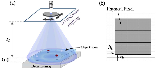
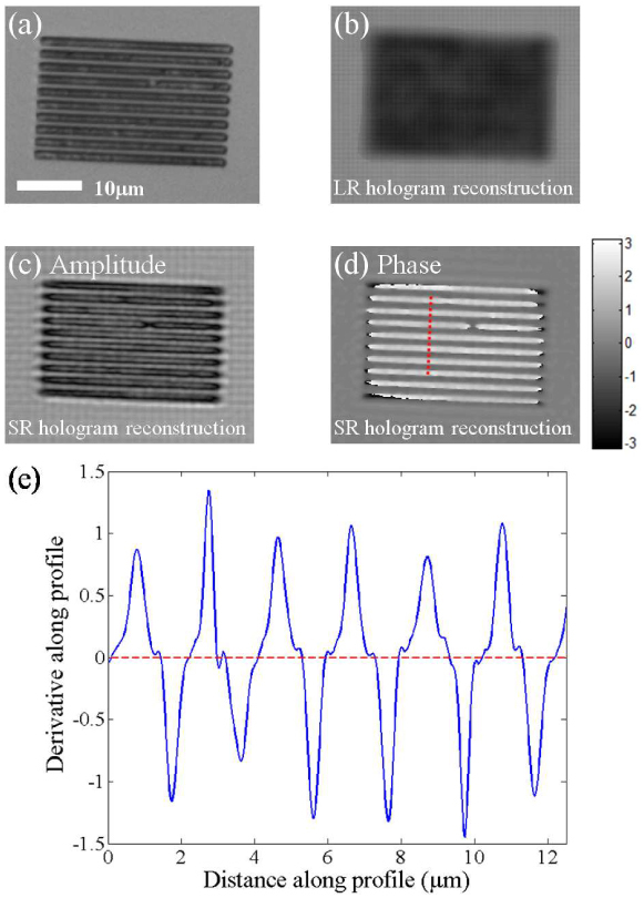
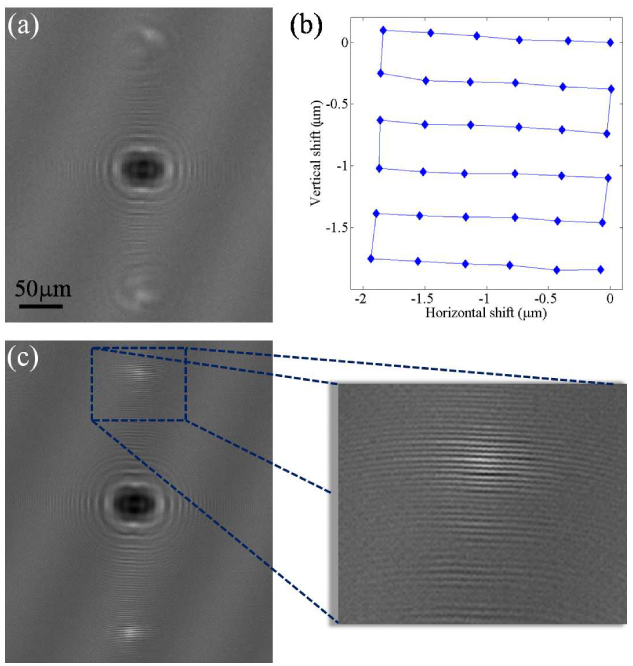
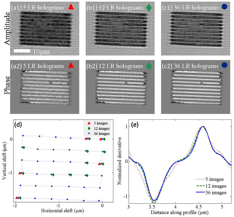
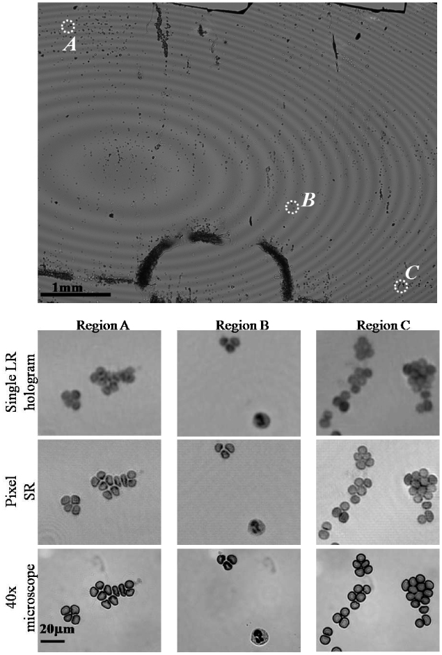
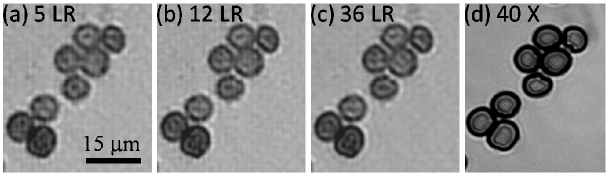
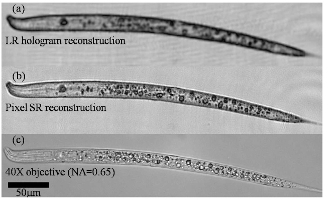
We demonstrate lensfree holographic microscopy on a chip to achieve approximately 0.6 microm spatial resolution corresponding to a numerical aperture of approximately 0.5 over a large field-of-view of approximately 24 mm2. By using partially coherent illumination from a large aperture (approximately 50 microm), we acquire lower resolution lensfree in-line holograms of the objects with unit fringe magnification. For each lensfree hologram, the pixel size at the sensor chip limits the spatial resolution of the reconstructed image. To circumvent this limitation, we implement a sub-pixel shifting based super-resolution algorithm to effectively recover much higher resolution digital holograms of the objects, permitting sub-micron spatial resolution to be achieved across the entire sensor chip active area, which is also equivalent to the imaging field-of-view (24 mm2) due to unit magnification. We demonstrate the success of this pixel super-resolution approach by imaging patterned transparent substrates, blood smear samples, as well as Caenoharbditis Elegans.


Figures

Fig. 1
Fig. 1
(a) Schematic diagram of our experimental setup. The aperture to object distance is much larger than the object to detector distance (z1~10 cm, z2<1mm). A shift of the aperture causes a demagnified shift of the object hologram formed at the detector plane, allowing sub-pixel hologram shifting. (b) Physical pixels captured in a single frame, here marked by bold borders, over imposed on the high-resolution pixel grid. This frame is shifted a distance of hk horizontally and vk vertically with respect to a reference frame.
Fig. 3
Fig. 3
(a) Microscope image of the object captured with a 40X objective lens (NA=0.65). (b) Amplitude reconstruction of the object using a single low-resolution hologram (see Fig. 2(a)). (c) Object amplitude reconstruction using the high-resolution hologram (see Fig. 2(c)) obtained from Pixel SR using 36 LR images. (d) Object phase reconstruction obtained from the same high-resolution hologram using Pixel SR. The object phase appears mostly positive due to phase wrapping. (e) The spatial derivative of the phase profile along the dashed line in pane (d). As explained in the text, this spatial derivative operation yields a train of delta functions with alternating signs, broadened by the PSF, which sets the resolution.
Fig. 2
Fig. 2
Multiple sub-pixel shifted lower-resolution holograms of the grating object are captured. One such lower-resolution hologram is shown in (a). The sub-pixel shifts between different holograms are automatically computed from the raw data using an iterative gradient method, the results of which are shown in (b). The Pixel SR algorithm recovers the high-resolution hologram of the object as shown in (c). The magnified portion of this super-resolution hologram shows high frequency fringes which were not captured in the lower-resolution holograms.
Fig. 4
Fig. 4
Comparison of pixel SR results using different number of LR holograms. Panes (a1-a2), (b1-b2), and (c1-c2) show the reconstructed amplitude and phase images of the same object using 5, 12, and 36 LR holograms, respectively. In (d), the sub-pixel shifts of the randomly chosen subsets of LR holograms are shown. In (e), the normalized spatial derivative profiles of the recovered phase images for each case (a2, b2 and c2) are shown, similar to Fig. 3(e).
Fig. 5
Fig. 5
Wide-field (FOV~24 mm2) high-resolution imaging of a whole blood smear sample using Pixel SR. A comparison among the image recovered using a single LR hologram (NA<0.2), the image recovered using Pixel SR (NA~0.5), and a 40X microscope image (NA=0.65) is provided for three regions of interest at different positions within the imaging FOV. Regions (A) and (C) show red blood cell clusters that are difficult to resolve using a single LR hologram, which are now clearly resolved using Pixel SR. In region (B) the sub-cellular features of a white blood cell are also resolved.
Fig. 6
Fig. 6
Similar to Fig. 4, we illustrate the pixel SR results of a red blood cell cluster achieved by using (a) 5 LR, (b) 12 LR and (c) 36 LR holograms. Following the same trend as in Fig. 4, almost the same reconstruction quality (especially in terms of the physical gaps among the cells) is achieved by feeding a sub-set of LR holograms to the pixel SR algorithm. (d) shows a 40X objective lens image of the same field of view acquired with NA=0.65.
Fig. 7
Fig. 7
Pixel super-resolution applied to imaging of C. elegans. (a) Recovered amplitude image from a single LR hologram. (b) Pixel SR image recovered using 16 sub-pixel shifted holograms. (c) Microscope image of the same worm captured with a 40X objective-lens (NA=0.65).

References

    1. Haddad W., Cullen D., Solem H., Longworth J., McPherson A., Boyer K., Rhodes C., “Fourier-transform holographic microscopy,” Appl. Opt. 31(24), 4973–4978 (1992).10.1364/AO.31.004973 - DOI - PubMed
    1. Schnars U., Jüptner W., “Direct recording of holograms by a CCD target and numerical reconstruction,” Appl. Opt. 33(2), 179–181 (1994).10.1364/AO.33.000179 - DOI - PubMed
    1. Zhang T., Yamaguchi I., “Three-dimensional microscopy with phase-shifting digital holography,” Opt. Lett. 23(15), 1221–1223 (1998).10.1364/OL.23.001221 - DOI - PubMed
    1. Cuche E., Bevilacqua F., Depeursinge C., “Digital holography for quantitative phase-contrast imaging,” Opt. Lett. 24(5), 291–293 (1999).10.1364/OL.24.000291 - DOI - PubMed
    1. Wagner C., Seebacher S., Osten W., Jüptner W., “Digital recording and numerical reconstruction of lensless fourier holograms in optical metrology,” Appl. Opt. 38(22), 4812–4820 (1999).10.1364/AO.38.004812 - DOI - PubMed

Publication types