

Real-time 4D signal processing and visualization using graphics processing unit on a regular nonlinear-k Fourier-domain OCT system
- PMID: 20589038
- PMCID: PMC2897754
- DOI: 10.1364/OE.18.011772
Real-time 4D signal processing and visualization using graphics processing unit on a regular nonlinear-k Fourier-domain OCT system
Abstract
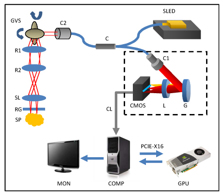
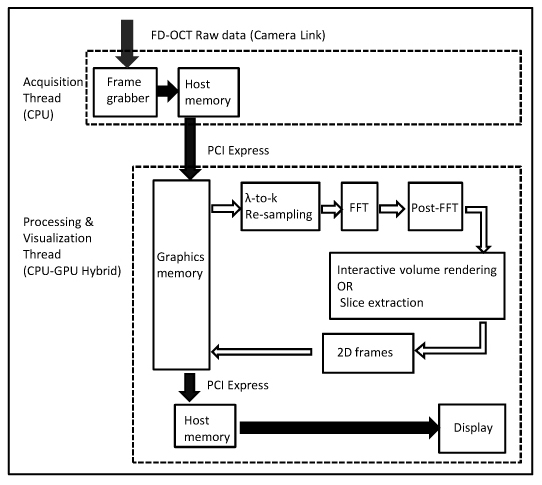
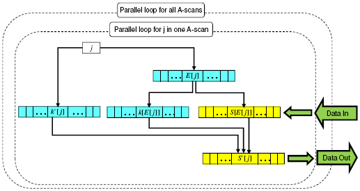
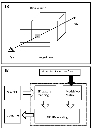
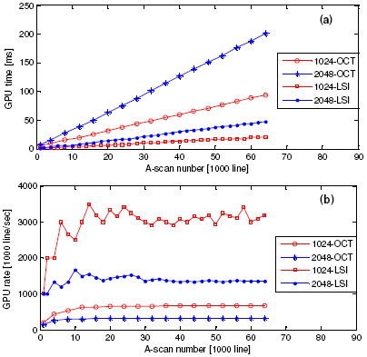
We realized graphics processing unit (GPU) based real-time 4D (3D+time) signal processing and visualization on a regular Fourier-domain optical coherence tomography (FD-OCT) system with a nonlinear k-space spectrometer. An ultra-high speed linear spline interpolation (LSI) method for lambda-to-k spectral re-sampling is implemented in the GPU architecture, which gives average interpolation speeds of >3,000,000 line/s for 1024-pixel OCT (1024-OCT) and >1,400,000 line/s for 2048-pixel OCT (2048-OCT). The complete FD-OCT signal processing including lambda-to-k spectral re-sampling, fast Fourier transform (FFT) and post-FFT processing have all been implemented on a GPU. The maximum complete A-scan processing speeds are investigated to be 680,000 line/s for 1024-OCT and 320,000 line/s for 2048-OCT, which correspond to 1GByte processing bandwidth. In our experiment, a 2048-pixel CMOS camera running up to 70 kHz is used as an acquisition device. Therefore the actual imaging speed is camera- limited to 128,000 line/s for 1024-OCT or 70,000 line/s for 2048-OCT. 3D Data sets are continuously acquired in real time at 1024-OCT mode, immediately processed and visualized as high as 10 volumes/second (12,500 A-scans/volume) by either en face slice extraction or ray-casting based volume rendering from 3D texture mapped in graphics memory. For standard FD-OCT systems, a GPU is the only additional hardware needed to realize this improvement and no optical modification is needed. This technique is highly cost-effective and can be easily integrated into most ultrahigh speed FD-OCT systems to overcome the 3D data processing and visualization bottlenecks.
Figures
Similar articles
-
Processing and rendering of Fourier domain optical coherence tomography images at a line rate over 524 kHz using a graphics processing unit.J Biomed Opt. 2011 Feb;16(2):020505. doi: 10.1117/1.3548153. J Biomed Opt. 2011. PMID: 21361661
-
Real time processing of Fourier domain optical coherence tomography with fixed-pattern noise removal by partial median subtraction using a graphics processing unit.J Biomed Opt. 2012 May;17(5):050503. doi: 10.1117/1.JBO.17.5.050503. J Biomed Opt. 2012. PMID: 22612118
-
Graphics processing unit accelerated non-uniform fast Fourier transform for ultrahigh-speed, real-time Fourier-domain OCT.Opt Express. 2010 Oct 25;18(22):23472-87. doi: 10.1364/OE.18.023472. Opt Express. 2010. PMID: 21164690 Free PMC article.
-
Medical image processing on the GPU - past, present and future.Med Image Anal. 2013 Dec;17(8):1073-94. doi: 10.1016/j.media.2013.05.008. Epub 2013 Jun 5. Med Image Anal. 2013. PMID: 23906631 Review.
-
An introductory tour of interactive rendering.IEEE Comput Graph Appl. 2006 Jan-Feb;26(1):76-87. doi: 10.1109/mcg.2006.9. IEEE Comput Graph Appl. 2006. PMID: 16463481 Review. No abstract available.
Cited by
-
Real-time 3D and 4D Fourier domain Doppler optical coherence tomography based on dual graphics processing units.Biomed Opt Express. 2012 Sep 1;3(9):2162-74. doi: 10.1364/BOE.3.002162. Epub 2012 Aug 20. Biomed Opt Express. 2012. PMID: 23024910 Free PMC article.
-
Real-time speckle variance swept-source optical coherence tomography using a graphics processing unit.Biomed Opt Express. 2012 Jul 1;3(7):1557-64. doi: 10.1364/BOE.3.001557. Epub 2012 Jun 7. Biomed Opt Express. 2012. PMID: 22808428 Free PMC article.
-
3D visualization of aqueous humor outflow structures in-situ in humans.Exp Eye Res. 2011 Sep;93(3):308-15. doi: 10.1016/j.exer.2011.03.019. Epub 2011 Apr 15. Exp Eye Res. 2011. PMID: 21514296 Free PMC article.
-
Morphometric analysis of aqueous humor outflow structures with spectral-domain optical coherence tomography.Invest Ophthalmol Vis Sci. 2012 Aug 7;53(9):5198-207. doi: 10.1167/iovs.11-9229. Invest Ophthalmol Vis Sci. 2012. PMID: 22499987 Free PMC article.
-
Automated non-rigid registration and mosaicing for robust imaging of distinct retinal capillary beds using speckle variance optical coherence tomography.Biomed Opt Express. 2013 May 7;4(6):803-21. doi: 10.1364/BOE.4.000803. Print 2013 Jun 1. Biomed Opt Express. 2013. PMID: 23761845 Free PMC article.
References
-
- Potsaid B., Gorczynska I., Srinivasan V. J., Chen Y., Jiang J., Cable A., Fujimoto J. G., “Ultrahigh speed spectral / Fourier domain OCT ophthalmic imaging at 70,000 to 312,500 axial scans per second,” Opt. Express 16(19), 15149–15169 (2008), http://www.opticsinfobase.org/oe/abstract.cfm?uri=oe-16-19-1514910.1364/OE.16.015149 - DOI - PMC - PubMed
-
- Grulkowski I., Gora M., Szkulmowski M., Gorczynska I., Szlag D., Marcos S., Kowalczyk A., Wojtkowski M., “Anterior segment imaging with Spectral OCT system using a high-speed CMOS camera,” Opt. Express 17(6), 4842–4858 (2009), http://www.opticsinfobase.org/oe/abstract.cfm?uri=oe-17-6-484210.1364/OE.17.004842 - DOI - PubMed
-
- Gora M., Karnowski K., Szkulmowski M., Kaluzny B. J., Huber R., Kowalczyk A., Wojtkowski M., “Ultra high-speed swept source OCT imaging of the anterior segment of human eye at 200 kHz with adjustable imaging range,” Opt. Express 17(17), 14880–14894 (2009), http://www.opticsinfobase.org/oe/abstract.cfm?uri=oe-17-17-1488010.1364/OE.17.014880 - DOI - PubMed
-
- Gargesha M., Jenkins M. W., Wilson D. L., Rollins A. M., “High temporal resolution OCT using image-based retrospective gating,” Opt. Express 17(13), 10786–10799 (2009), http://www.opticsinfobase.org/oe/abstract.cfm?uri=oe-17-13-1078610.1364/OE.17.010786 - DOI - PMC - PubMed
Publication types
MeSH terms
Grants and funding
LinkOut - more resources
Full Text Sources
Other Literature Sources
Research Materials
