. 2010 Jun 10:4:15.
doi: 10.3389/fnsys.2010.00015. eCollection 2010.

Different corticostriatal integration in spiny projection neurons from direct and indirect pathways

Affiliations

Different corticostriatal integration in spiny projection neurons from direct and indirect pathways

Edén Flores-Barrera et al. Front Syst Neurosci. .

Abstract

The striatum is the principal input structure of the basal ganglia. Major glutamatergic afferents to the striatum come from the cerebral cortex and make monosynaptic contacts with medium spiny projection neurons (MSNs) and interneurons. Also: glutamatergic afferents to the striatum come from the thalamus. Despite differences in axonal projections, dopamine (DA) receptors expression and differences in excitability between MSNs from "direct" and "indirect" basal ganglia pathways, these neuronal classes have been thought as electrophysiologically very similar. Based on work with bacterial artificial chromosome (BAC) transgenic mice, here it is shown that corticostriatal responses in D(1)- and D(2)-receptor expressing MSNs (D(1)- and D(2)-MSNs) are radically different so as to establish an electrophysiological footprint that readily differentiates between them. Experiments in BAC mice allowed us to predict, with high probability (P > 0.9), in rats or non-BAC mice, whether a recorded neuron, from rat or mouse, was going to be substance P or enkephalin (ENK) immunoreactive. Responses are more prolonged and evoke more action potentials in D(1)-MSNs, while they are briefer and exhibit intrinsic autoregenerative responses in D(2)-MSNs. A main cause for these differences was the interaction of intrinsic properties with the inhibitory contribution in each response. Inhibition always depressed corticostriatal depolarization in D(2)-MSNs, while it helped in sustaining prolonged depolarizations in D(1)-MSNs, in spite of depressing early discharge. Corticostriatal responses changed dramatically after striatal DA depletion in 6-hydroxy-dopamine (6-OHDA) lesioned animals: a response reduction was seen in substance P (SP)+ MSNs whereas an enhanced response was seen in ENK+ MSNs. The end result was that differences in the responses were greatly diminished after DA depletion.

Keywords: 6-hydroxy-dopamine; GABAA receptors; Parkinson disease; basal ganglia; corticostriatal pathway; medium spiny neurons; strionigral pathway; striopallidal pathway.


Figures
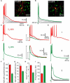
Figure 1
Figure 1
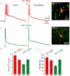
Different corticostriatal response in D1 or D2 eGFP medium spiny neurons (MSNs) from BAC mice. (A,B) Families of subthreshold and suprathreshold corticostriatal synaptic responses for representative D1- (red traces) and D2- (green traces) MSNs obtained from BAC transgenic mice striatum. Insets show biocytin-filled recorded cells and green fluorescent protein positive (GFP+) accompanying cells (insets). Yellow color of recorded cells indicate D1- (A) and D2-eGFP-positive cells (B), respectively (superimposition). Notice that D1-MSNs suprathreshold response is more prolonged and generates more action potentials whereas D2-MSNs response only display an initial burst of high frequency firing. (C,D) Area under response is smaller in D2-MSNs, as well as half-widths (F–H) Histograms summarize measurements in neuronal samples (half-widths and areas under responses): n = 55 D1-MSNs and n = 50 D2-MSNs. Differences were significant. (E) Traces at a slower sweep (1–3) illustrate frequency of discharge in D1- (1), D2-MSNs (2) as well as intrinsic autoregenerative responses in D2-MSNs. (I) Firing frequency was higher in D2-MSNs.
Figure 2
Figure 2
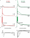
Contribution of synaptic inhibition differs in corticostriatal responses from D1- and D2-MSNs from BAC mice. (A,E) Suprathreshold corticostriatal responses in D1- and D2-MSNs, respectively. (B,F) Addition of 10 μM bicuculline to the bath saline (colored traces) changes both responses indicating that GABAergic events participate in each of them. (C,G) Superimposed records: (A) with (B) and (E) with (F), show that GABAergic blockade mostly reduce D1-MSNs response and enhance D2-MSNs response, respectively. Notice prolongation of action potentials trains in D2-MSNs with inactivation of some spikes. (D,H) Digital subtraction of records in (C), (G) disclose the bicuculline-sensitive components in both neuron classes. Notice that except for an initial restrain in evoked discharge, the bicuculline-sensitive component depolarizes D1-MSNs. Insets: histograms show that bicuculline first enhances, then depresses D1-MSNs response (a biphasic response, red bars, n = 24). In contrast, bicuculline only augments D2-MSNs response (green bar, n = 22). In the same way, half-widths were decreased and enhanced for D1- and D2 MSNs, respectively.
Figure 3
Figure 3
Bicuculline actions differ in D1- and D2-MSNs at different stimulation intensities. (A,E) Blockade of GABAergic synapses during subthreshold synaptic responses (black trace = control; colored trace = after bicuculline): both responses are depolarizing and have a similar amplitude in control conditions (about 10 mV). Notice that GABAergic blockade reduces the synaptic response in D1- and increases the synaptic response in D2-MSNs. There is no hyperpolarizing synaptic component in any response in spite of both being bicuculline-sensitive: inhibition is depolarizing in both cases. (B,F) Blockade of GABAergic synapses during threshold synaptic responses. Again, a decrease and an increase in the responses were observed in D1- and D2-MSNs synaptic responses, respectively. Notice that blockade of GABAergic synapses impedes the firing of action potentials in D1-MSNs (B) and elicits a local response that distorts synaptic kinetics in D2-MSNs (F). (C,G) Blockade of GABAergic synapses in suprathreshold synaptic responses. Superimposed traces cross each other in D1-MSNs and do not cross in D2-MSNs. (D–H) Digital subtractions of traces in (AG): Bicuculline-sensitive component helps in depolarizing all D1-MSNs synaptic responses, except at the beginning of the suprathreshold responses. In contrast, bicuculline enhanced D2-MSNs synaptic responses at all stimulus strengths. Inhibition is depolarizing and even excitatory in D1-MSNs whereas it always represses D2-MSNs (shunting inhibition).
Figure 4
Figure 4
Action of an inverse selective agonist of GABAA receptors containing the α5 subunit on D1- and D2-MSNs corticostriatal responses. (A) Superimposed corticostriatal responses from a D1-MSN in control (black) and during 10 μM L655-708 (red). (B) Superimposed corticostriatal responses from a D2-MSN in control (black) and during 10 μM L655-708 (green). (C) Histogram shows that L655-708 affected both responses similarly (n = 4).
Figure 5
Figure 5
Corticostriatal synaptic responses of SP+ MSNs and ENK+ MSNs as modified by unilateral 6-OHDA lesions in the SNc. (A) Representative suprathreshold corticostriatal response of a SP+ MSN recorded from an intact rat. (B) Representative suprathreshold corticostriatal response of a SP+ MSN recorded in a 6-OHDA lesioned rat. Notice a response decreased in both amplitude and duration; the train of action potentials is substituted by a brief burst. (C) Representative suprathreshold corticostriatal response of an ENK+ MSN from an intact rat. (D) Representative suprathreshold corticostriatal response of an ENK+ MSN recorded in a 6-OHDA lesioned rat. Notice enhanced depolarization. Inset shows double staining of the recorded and biocytin-filled SP+ and ENK+ MSNs. (E) Histograms of areas under synaptic responses in intact (ctl) and DA-depleted (-DA) samples of neurons. Notice a decrease in the area of SP+ MSNs responses and an increase in the area of ENK+ MSNs responses. After DA depletion areas from both neuronal responses are not significantly different. (F) Histograms of half-widths of corticostriatal responses in intact (ctl) and DA-depleted (-DA) neurons. Response half-widths decreased in SP+ MSNs and increased in ENK+ MSNs. SP+ MSNs (red): n = 11 in control and n = 16 after DA depletion. ENK+ MSNs (green): n = 9 in control and n = 13 after DA depletion. Differences were significant when comparing control and DA-depleted neurons of the same class, and when comparing control responses between neuronal classes. Differences became non-significant when comparing neuronal classes after DA depletion.
Figure 6
Figure 6
Comparison of corticostriatal responses evoked by different stimulus intensities in SP+ and ENK+ MSNs from intact and lesioned (6-OHDA) animals. (A–C) Superimposition of control, (black traces) and DA-depleted (red traces), subthreshold, threshold – spikes clipped – and suprathreshold corticostriatal responses in a representative SP+ MSN. Except for the suprathreshold case, influence of DA depletion in the responses is hardly noticed. In the suprathreshold response depolarizing inhibition is decreased after DA depletion; similarly to the action of bicuculline (cf., Figure 3). (D–F) Superimposition of control, (black traces) and DA-depleted (green traces), subthreshold, threshold – spikes clipped – and suprathreshold corticostriatal responses in a representative ENK+ MSN. Notice that the responses are more depolarized and prolonged than their corresponding controls modifying the kinetics of purely synaptic inputs. (G–I) Superimposition of corticostriatal responses from SP+ (red) and an ENK+ (green) MSNs after DA depletion: notice smaller and less prolonged responses for SP+ MSNs, except for the suprathreshold responses that look similar.
Figure 7
Figure 7
Actions of bicuculline on corticostriatal responses are modified after DA depletion. (A,E) Subthreshold synaptic responses of SP+ and ENK+ neurons depleted of DA (black traces) are compared. Both responses are depolarizing in spite of containing inhibitory components as demonstrated by the actions of 10 μM bicuculline (colored traces). Bicuculline reduces SP+ MSNs response whereas it enhances ENK+ MSNs synaptic responses. However, enhancing bicuculline action on ENK+ MSNs is larger than in intact animals (cf., Figure 3) exhibiting a local response that distorts the kinetics of the evoked synaptic event. (B,F) Threshold synaptic responses after DA depletion (black traces) from the same neurons are compared before and during bicuculline (colored traces): Notice absence of bicuculline action in SP+ MSNs at threshold level, suggesting a decrease in inhibitory inputs. In the case of ENK+ MSNs, an intrinsic depolarization that deforms synaptic kinetics is disclosed. (C,G) Suprathreshold corticostriatal responses after DA depletion (black traces) are compared in the same neurons before and during bicuculline (colored traces): a great decrease in bicuculline action is clearly visible in SP+ MSNs (cf., Figure 3), confirming that recruiting of inhibitory inputs is hindered after attaining certain stimulus strengths. A robust depolarization with a prolonged train of spikes is evident in ENK+ MSNs at suprathreshold levels. (D) Digital subtractions of responses in SP+ MSNs (a–c) are signaled by corresponding letters. Notice: initial depolarization due to the inhibitory component is much decreased in subthreshold responses (a), while at threshold level bicuculline action seems occluded or not participating (b). Suprathreshold response reveals inhibition of initial depolarization (c) but an almost complete lack of inhibitory participation in the sustaining of the late depolarizing plateau potential. (H) By comparison, bicuculline-sensitive components subtracted from ENK+ MSNs responses (e–g) have about the same amplitude for all responses, although more prolonged with increases in stimulus strength, suggesting the activation of intrinsic currents.

Similar articles

Cited by

References

    1. Ade K. K., Janssen M. J., Ortinski P. I., Vicini S. (2008). Differential tonic GABA conductances in striatal medium spiny neurons. J. Neurosci. 28, 1185–119710.1523/JNEUROSCI.3908-07.2008 - DOI - PMC - PubMed
    1. Bargas J., Galarraga E., Aceves J. (1991). Dendritic activity on neostriatal neurons as inferred from somatic intracellular recordings. Brain Res. 539, 159–16310.1016/0006-8993(91)90700-6 - DOI - PubMed
    1. Bartos M., Vida I., Jonas P. (2007). Synaptic mechanisms of synchronized gamma oscillations in inhibitory interneuron networks. Nat. Rev. Neurosci. 8, 45–5610.1038/nrn2044 - DOI - PubMed
    1. Bennett B. D., Bolam J. P. (1994). Synaptic input and output of parvalbumin-immunoreactive neurons in the neostriatum of the rat. Neuroscience 62, 707–71910.1016/0306-4522(94)90471-5 - DOI - PubMed
    1. Bracci E., Panzeri S. (2006). Excitatory GABAergic effects in striatal projection neurons. J. Neurophysiol. 95, 1285–129010.1152/jn.00598.2005 - DOI - PubMed

LinkOut - more resources