. 2011 Jun;10(3):295-306.
doi: 10.1007/s10237-010-0235-5. Epub 2010 Jun 30.

Models of cardiac electromechanics based on individual hearts imaging data: image-based electromechanical models of the heart

Affiliations

Models of cardiac electromechanics based on individual hearts imaging data: image-based electromechanical models of the heart

Viatcheslav Gurev et al. Biomech Model Mechanobiol. 2011 Jun.

Erratum in

  • Biomech Model Mechanobiol. 2011 Jun;10(3):307

Abstract

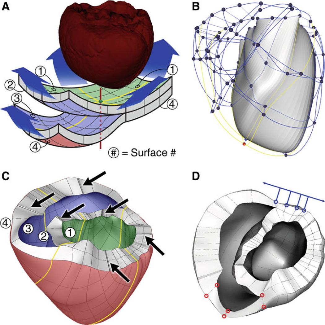
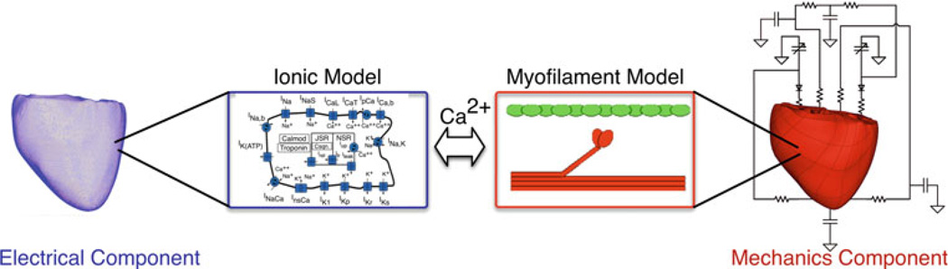
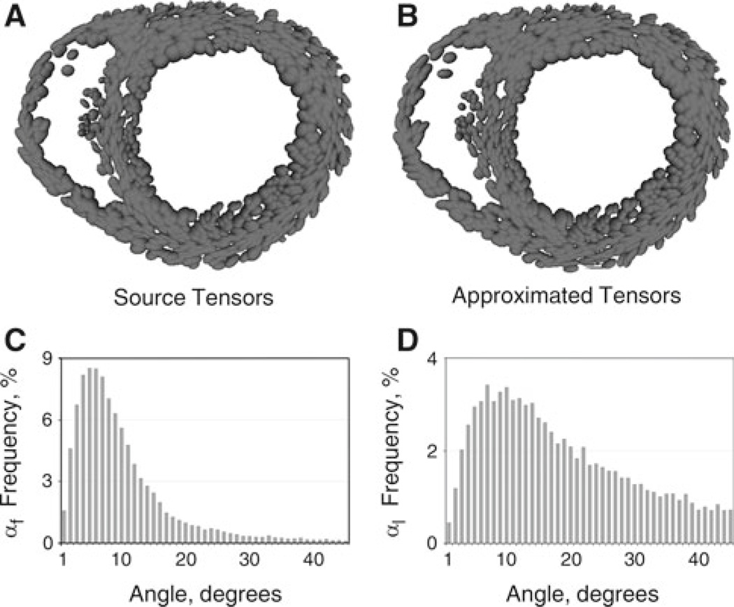
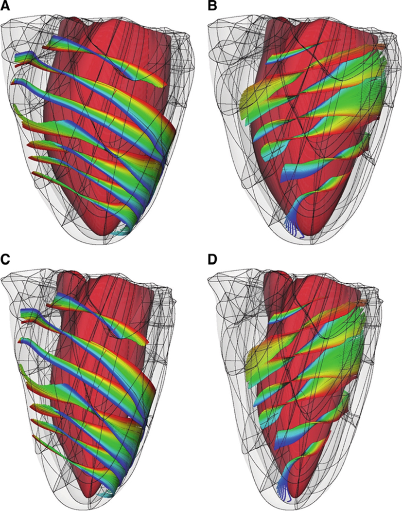
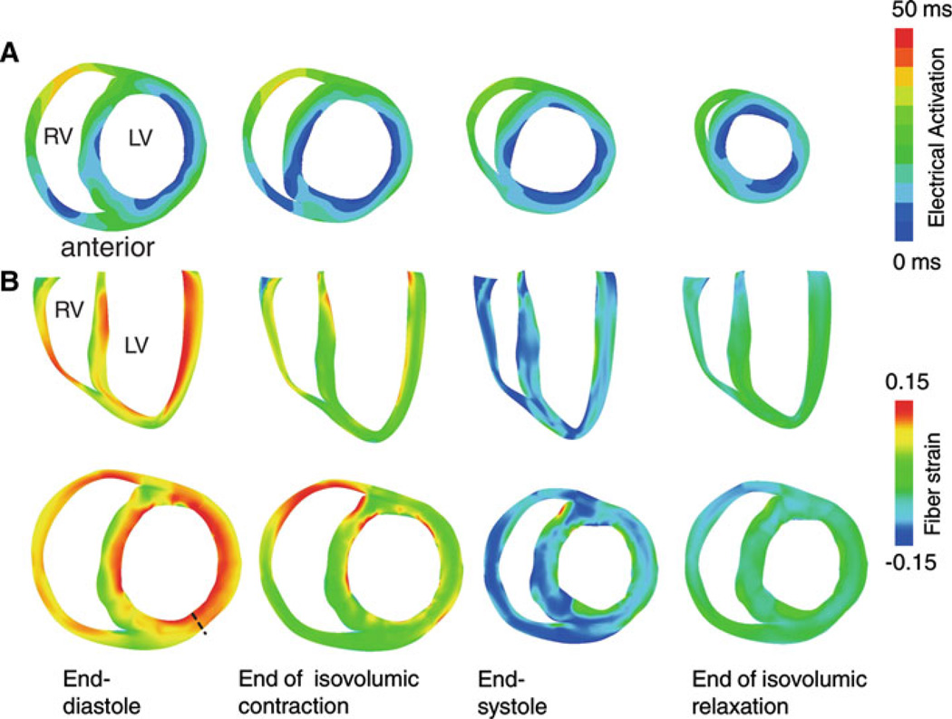
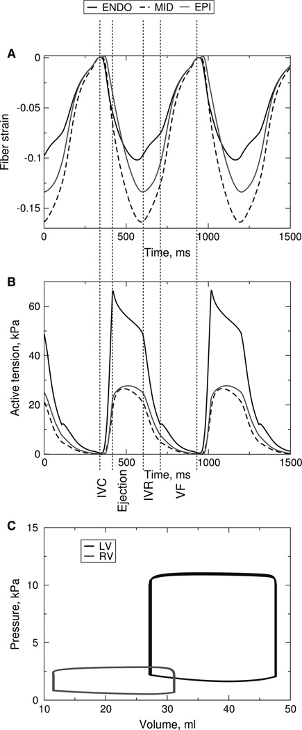
Current multi-scale computational models of ventricular electromechanics describe the full process of cardiac contraction on both the micro- and macro- scales including: the depolarization of cardiac cells, the release of calcium from intracellular stores, tension generation by cardiac myofilaments, and mechanical contraction of the whole heart. Such models are used to reveal basic mechanisms of cardiac contraction as well as the mechanisms of cardiac dysfunction in disease conditions. In this paper, we present a methodology to construct finite element electromechanical models of ventricular contraction with anatomically accurate ventricular geometry based on magnetic resonance and diffusion tensor magnetic resonance imaging of the heart. The electromechanical model couples detailed representations of the cardiac cell membrane, cardiac myofilament dynamics, electrical impulse propagation, ventricular contraction, and circulation to simulate the electrical and mechanical activity of the ventricles. The utility of the model is demonstrated in an example simulation of contraction during sinus rhythm using a model of the normal canine ventricles.


Figures

Fig. 1
Fig. 1
a Fitting the mechanics mesh to the ventricular geometry obtained from segmenting the MR imaging stacks. b The initial mechanics mesh. The LV is solid and the RV is transparent. The yellow lines correspond to those in panel A. c Final mechanics mesh. The arrows point to locations where corner elements were removed. d Locations at which boundary conditions were applied. See text for details
Fig. 2
Fig. 2
Schematic diagram of the electromechanics model
Fig. 3
Fig. 3
Anterior view of the finite element mechanics meshes. From left to right: normal canine, failing canine, and human ventricles
Fig. 4
Fig. 4
Tensor interpolation and comparison with the original DTMR imaging data for the normal canine ventricles. Tensors from DTMR images (a) and approximated tensors (b) are represented as ellipses, where the lengths of each axis correspond to the DT eigenvalues. Frequency distribution bar graphs of the angles αf (c) and αl (d)
Fig. 5
Fig. 5
Fiber architecture of the normal canine ventricles at end-diastole (a, b) and end-systole (c, d). For clarity of visualization, fibers near the epicardium (a, c) and near the endocardium (b, d) are shown separately. The colors trace individual fibers
Fig. 6
Fig. 6
Electrical activation and transmural fiber strain during sinus rhythm. a Isochronal maps of electrical activation in a short-axis view during sinus rhythm. b Transmural fiber strain during the cardiac cycle in long-axis (top) and short-axis (bottom) views. The undeformed (stress-free) state was the reference state for strain calculation
Fig. 7
Fig. 7
a Fiber strain for different wall depths at the mid-base of the anterior left ventricular wall. The end-diastolic state is the reference state for the strain calculations. b Active tensions at the same locations as in panel A. c LV and RV pressure-volume loops during the cardiac cycles. IVC: isovolumic contraction, IVR: isovolumic relaxation, VF: ventricular filling

References

    1. Arsigny V, Fillard P, Pennec X, Ayache N. Log-Euclidean metrics for fast and simple calculus on diffusion tensors. Magn Reson Med. 2006;56:411–421. - PubMed
    1. Arts T, Reneman R, Veenstra P. A model of the mechanics of the left ventricle. Ann Biomed Eng. 1979;7:299–318. - PubMed
    1. Ashikaga H, Coppola B, Hopenfeld B, Leifer E, McVeigh E, Omens J. Transmural dispersion of myofiber mechanics: implications for electrical heterogeneity in vivo. J Am Coll Cardiol. 2007;49:909–916. - PMC - PubMed
    1. Ashikaga H, Omens JH, Ingels NB, Covell JW. Transmural mechanics at left ventricular epicardial pacing site. Am J Physiol Heart Circ Physiol. 2004;286:H2401–H2407. - PMC - PubMed
    1. Balay S, Buschelman K, Eijkhout V, Cropp W, Kaushik D, Knepley M, McInnes LC, Smith B, Zhang H. PETSc User manual, vol ANL-95/11-Revision 2.3.3. Argonne: Argonne National Laboratory; 2007.

Publication types

LinkOut - more resources