. 2010 Nov 1;107(4):683-95.
doi: 10.1002/bit.22850.

Three-dimensional culture systems for the expansion of pluripotent embryonic stem cells

Affiliations
Free PMC article

Three-dimensional culture systems for the expansion of pluripotent embryonic stem cells

Michael P Storm et al. Biotechnol Bioeng. .
Free PMC article

Abstract

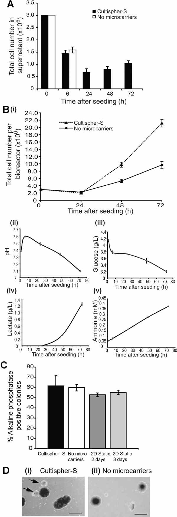
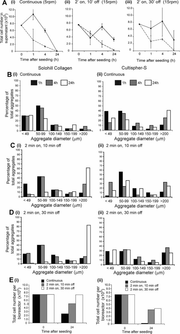
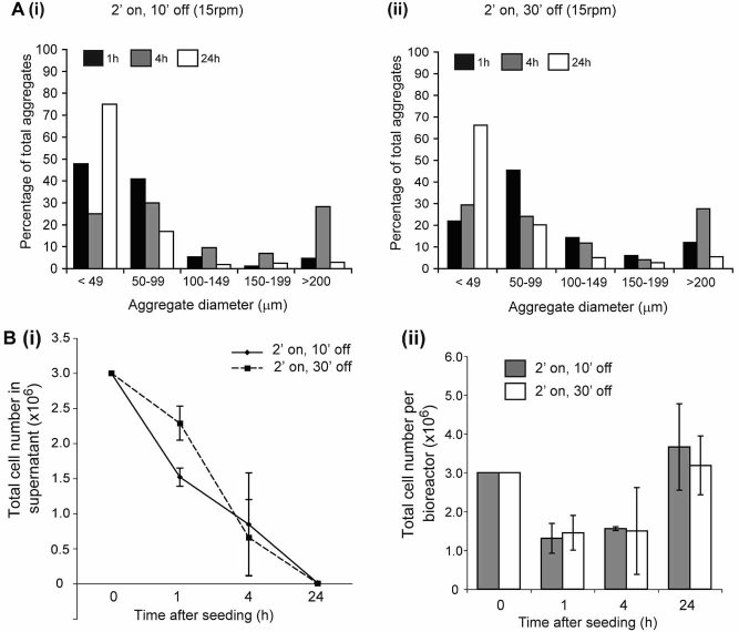
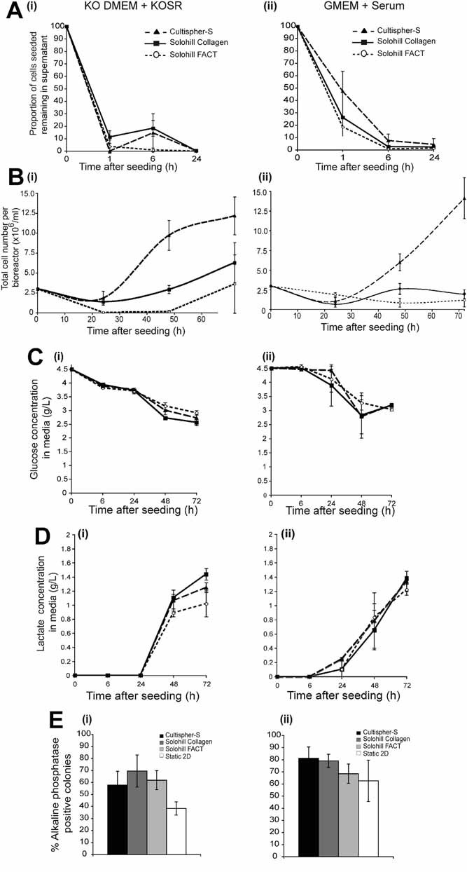
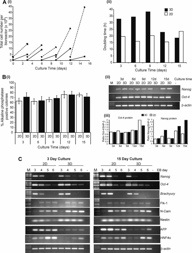
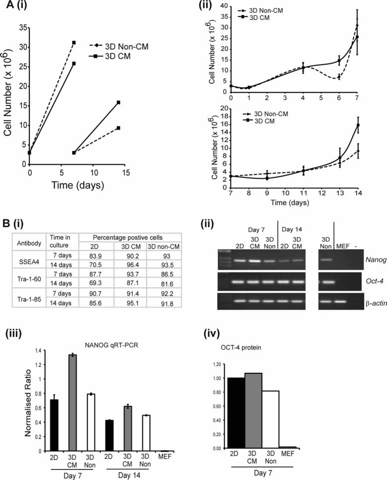
Mouse embryonic stem cell (ESC) lines, and more recently human ESC lines, have become valuable tools for studying early mammalian development. Increasing interest in ESCs and their differentiated progeny in drug discovery and as potential therapeutic agents has highlighted the fact that current two-dimensional (2D) static culturing techniques are inadequate for large-scale production. The culture of mammalian cells in three-dimensional (3D) agitated systems has been shown to overcome many of the restrictions of 2D and is therefore likely to be effective for ESC proliferation. Using murine ESCs as our initial model, we investigated the effectiveness of different 3D culture environments for the expansion of pluripotent ESCs. Solohill Collagen, Solohill FACT, and Cultispher-S microcarriers were employed and used in conjunction with stirred bioreactors. Initial seeding parameters, including cell number and agitation conditions, were found to be critical in promoting attachment to microcarriers and minimizing the size of aggregates formed. While all microcarriers supported the growth of undifferentiated mESCs, Cultispher-S out-performed the Solohill microcarriers. When cultured for successive passages on Cultispher-S microcarriers, mESCs maintained their pluripotency, demonstrated by self-renewal, expression of pluripotency markers and the ability to undergo multi-lineage differentiation. When these optimized conditions were applied to unweaned human ESCs, Cultispher-S microcarriers supported the growth of hESCs that retained expression of pluripotency markers including SSEA4, Tra-1-60, NANOG, and OCT-4. Our study highlights the importance of optimization of initial seeding parameters and provides proof-of-concept data demonstrating the utility of microcarriers and bioreactors for the expansion of hESCs.


Figures

Figure 1
Figure 1
Culture of mESCs on macroporous microcarriers supports their expansion. mESCs were seeded onto Cultispher-S microcarriers at 6 × 10⁴ cells/mL in KO-DMEM plus KO-SR and LIF or directly into bioreactors lacking microcarriers. Impeller speed was continuous at 15 rpm for 2 h and 60 rpm subsequently. A: Average number of live cells remaining in the supernatant are shown (n = 3, ±SEM). B: (i) Total number of viable cells attached to microcarriers (dashed line) or in aggregates (solid line) per bioreactor were determined and media was analyzed for (ii) pH, (iii) glucose, (iv) lactate, and (v) ammonia. Mean values are shown (n = 3, ±SEM). C: Following 72 h of bioreactor culture, self-renewal of mESCs was assessed and alkaline phosphatase positive colonies scored. Mean values are shown (n = 3, ±SEM). “2D Static” refers to E14 mESCs maintained on 2D-tissue culture plates for 2 or 3 days. D: Photographs were taken at 72 h of (i) Cultispher-S-containing cultures and (ii) cultures without microcarriers. Black arrows indicate cell aggregates formed in Cultispher-S cultures, which are smaller and more transparent than the microcarriers themselves and which can attach at a single point to the microcarriers. Scale bar = 200 µm.
Figure 2
Figure 2
Initial seeding parameters play a critical role in minimizing aggregate formation. A: mESCs were seeded in triplicate onto either Solohill Collagen (solid lines) or Cultispher-S (dashed line) microcarriers at a density of 1.5 × 10⁴ cells/mL in KO-DMEM plus KO-SR and LIF. Seeding was carried out for 24 h with (i) continuous stirring of 5 rpm, (ii) 2 min stirring at 15 rpm followed by 10 min off, or (iii) 2 min stirring at 15 rpm followed by 30 min off. The average number of cells (±SD) remaining in the supernatant are shown. B–D: The diameters of more than 200 cell aggregates were measured and the percentage of aggregates in each of the ranges indicated calculated for each time and condition. B: Continuous stirring of 5 rpm; C: 2 min at 15 rpm, 10 min off, and D: 2 min at 15 rpm, 30 min off. Samples B(i), C(i), and D(i) represent data for Solohill collagen, while B(ii), C(ii), and D(ii) represent data for Cultispher-S. E: The total number of viable cells recoverable from each bioreactor at 24 h was determined (i) Solohill collagen and (ii) Cultispher-S microcarriers.
Figure 3
Figure 3
Increased initial seeding density minimizes aggregate formation. A: mESCs were seeded onto Cultispher-S at a density of 6 × 10⁴ cells/mL using either (i) 2 min stirring at 15 rpm followed by 10 min off or (ii) 2 min stirring at 15 rpm followed by 30 min off. Samples were collected and the percentage of aggregates in the ranges indicated calculated for each condition. B: (i) The average number of cells remaining in the supernatant (n = 3, ±SD) and (ii) the total number of viable cells recoverable from each bioreactor (mean ± SD, n = 3) are shown.
Figure 4
Figure 4
Media of different compositions support expansion of mESCs on microcarriers in bioreactors. mESCs were seeded at 6 × 10⁴ cells/mL in either (i) KO-DMEM plus KO-SR and LIF or (ii) GMEM plus serum (10% v/v) and LIF, using 2 min at 15 rpm followed by 10 min off, onto each of three microcarriers in parallel in duplicate: Cultispher-S (dashed lines, filled triangle), Solohill collagen (solid lines, filled square), or Solohill FACT (dotted lines, open circles). A: The number of cells remaining in the supernatant were determined and the average proportion of cells seeded that remained in the supernatant (±SD) are shown. B: The mean numbers of viable cells attached to microcarriers (n = 3, ±SD) are shown for each condition. The mean levels (±SD) of (C) glucose and (D) lactate are shown. E: Self-renewal was assessed after 72 h as described in legend to Figure 1C.
Figure 5
Figure 5
mESCs cultivated long-term on microcarriers retain their undifferentiated state. mESC were seeded onto Cultispher-S microcarriers as described in the legend for Figure 4 (3D samples) or plated onto 10 cm tissue culture plates (2 × 10⁵ total) in the same culture medium (2D samples). Cells were passaged every 3 days and total cell numbers recovered were recorded prior to re-plating using the same conditions described above. A: (i) Cell numbers plated and recovered at each passage for each condition are presented. 2D samples are indicated by the solid line and 3D samples by the dashed line. (ii) Population doubling times for cells in 3D and 2D cultures are shown. B: At each passage cells were trypsined. (i) Self-renewal was assessed and alkaline phosphatase positive colonies scored. Means ± SD from two independent long-term culture experiments for each condition are shown. (ii) RNA or (iii) protein were extracted from cells cultured in 2D or 3D conditions and expression of Oct-4 and Nanog determined by semi-quantitative RT-PCR (ii) or quantitative immunoblotting (iii). C: mESCs cultured in 2D or 3D for 3 or 15 days were plated into methylcellulose to generate embryoid bodies. EBs were harvested, RNA extracted and expression of marker genes analyzed by semi-quantitative PCR. M, molecular markers; −, PCR negative control.
Figure 6
Figure 6
Cultispher-S microcarriers support expansion of undifferentiated SHEF3 hESCs. 3 × 10⁶ SHEF3 hESCs were seeded onto Cultispher-S microcarriers in KO-DMEM supplemented with 20% (v/v) KO-SR and 4 ng/mL FGF-2 in the presence (3D CM) or absence (3D Non-CM) of MEF conditioned medium (CM). Cells were cultured for 7 days, passaged and then cultured for a further 7 days. A: (i) Cell numbers plated and recovered at each passage for each condition are presented. (ii) The mean number of viable cells attached to microcarriers (±SD) are shown for each condition for the culture periods d0–d7 (upper panel) and d7–d14 (lower panel). B: Assessment of markers of pluripotency. (i) Expression of SSEA4, Tra-1–60 and Tra-1–85, were determined by flow cytometry. 2D indicates hESCs controls maintained in 2D culture on MEF plus FGF-2. RNA and protein were extracted from hESCs cultured in 2D or 3D conditions for the times indicated and expression of OCT-4 and NANOG determined by (ii) semi-quantitative RT-PCR; (iii) quantitative RT-PCR normalized to β-actin (±SD) or (iv) immunoblotting, normalized to SHP-2.
Figure 7
Figure 7
Nutrient and metabolite profiles of SHEF3 hESC-Cultispher-S bioreactor cultures. SHEF3 hESCs were seeded onto Cultispher-S. Media samples were harvested and analyzed for the concentration of: (A) glucose; (B) lactate, and (C) ammonia. D: The pH of media samples. Triplicate measurements were taken from duplicate samples for MEF CM and Non-CM conditions during the first 7-day culture period, averages ± SD are shown. 2D samples are a single replicate. [Color figure can be viewed in the online issue, which is available at http://www.interscience.wiley.com.]

References

    1. Abranches E, Bekman E, Henrique D, Cabral JM. Expansion of mouse embryonic stem cells on microcarriers. Biotechnol Bioeng. 2007;96(6):1211–1221. - PubMed
    1. Bone HK, Welham MJ. Phosphoinositide 3-kinase signalling regulates early development and developmental haemopoiesis. J Cell Sci. 2007;120:1752–1762. - PMC - PubMed
    1. Cormier JT, zur Nieden NI, Rancourt DE, Kallos MS. Expansion of undifferentiated murine embryonic stem cells as aggregates in suspension culture bioreactors. Tissue Eng. 2006;12(11):3233–3245. - PubMed
    1. Dang SM, Gerecht-Nir S, Chen J, Itskovitz-Eldor J, Zandstra PW. Controlled, scalable embryonic stem cell differentiation culture. Stem Cells. 2004;22(3):275–282. - PubMed
    1. Draper JS, Smith K, Gokhale P, Moore HD, Maltby E, Johnson J, Meisner L, Zwaka TP, Thomson JA, Andrews PW. Recurrent gain of chromosomes 17q and 12 in cultured human embryonic stem cells. Nat Biotechnol. 2004;22(1):53–54. - PubMed

Publication types