

Three-dimensional structure of the weakly associated protein homodimer SeR13 using RDCs and paramagnetic surface mapping
- PMID: 20589905
- PMCID: PMC2975131
- DOI: 10.1002/pro.447
Three-dimensional structure of the weakly associated protein homodimer SeR13 using RDCs and paramagnetic surface mapping
Abstract
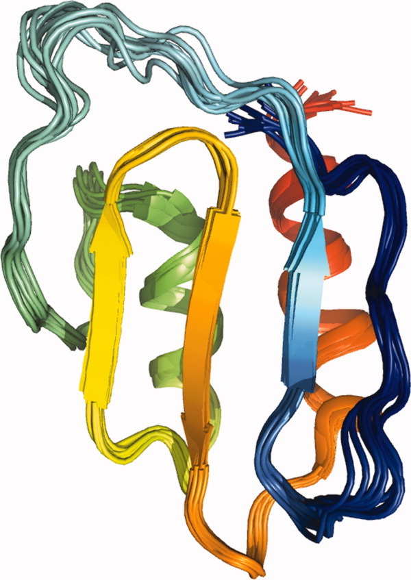
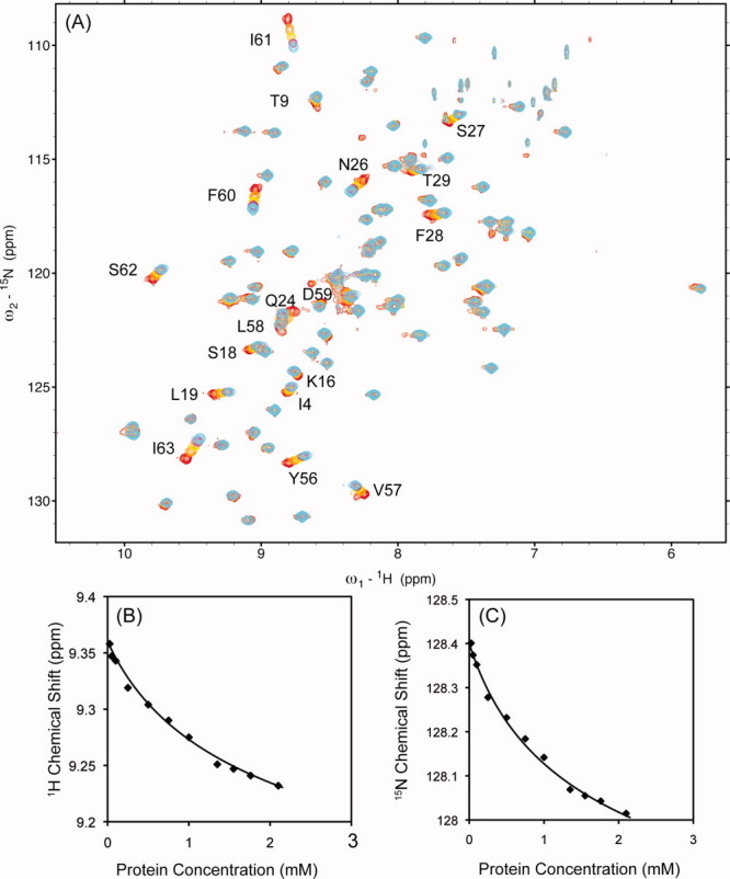
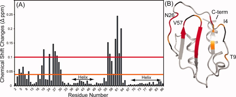
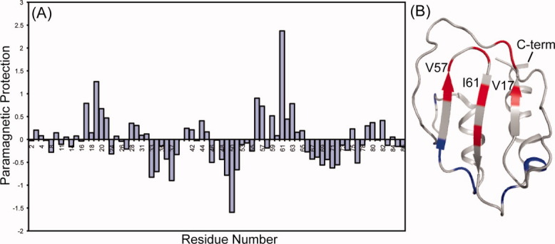
The traditional NMR-based method for determining oligomeric protein structure usually involves distinguishing and assigning intra- and intersubunit NOEs. This task becomes challenging when determining symmetric homo-dimer structures because NOE cross-peaks from a given pair of protons occur at the same position whether intra- or intersubunit in origin. While there are isotope-filtering strategies for distinguishing intra from intermolecular NOE interactions in these cases, they are laborious and often prove ineffectual in cases of weak dimers, where observation of intermolecular NOEs is rare. Here, we present an efficient procedure for weak dimer structure determination based on residual dipolar couplings (RDCs), chemical shift changes upon dilution, and paramagnetic surface perturbations. This procedure is applied to the Northeast Structural Genomics Consortium protein target, SeR13, a negatively charged Staphylococcus epidermidis dimeric protein (K(d) 3.4 ± 1.4 mM) composed of 86 amino acids. A structure determination for the monomeric form using traditional NMR methods is presented, followed by a dimer structure determination using docking under orientation constraints from RDCs data, and scoring under residue pair potentials and shape-based predictions of RDCs. Validation using paramagnetic surface perturbation and chemical shift perturbation data acquired on sample dilution is also presented. The general utility of the dimer structure determination procedure and the possible relevance of SeR13 dimer formation are discussed.
Copyright © 2010 The Protein Society.
Figures
Similar articles
-
Determination of the structures of symmetric protein oligomers from NMR chemical shifts and residual dipolar couplings.J Am Chem Soc. 2011 Apr 27;133(16):6288-98. doi: 10.1021/ja111318m. Epub 2011 Apr 5. J Am Chem Soc. 2011. PMID: 21466200 Free PMC article.
-
A graphical method for analyzing distance restraints using residual dipolar couplings for structure determination of symmetric protein homo-oligomers.Protein Sci. 2011 Jun;20(6):970-85. doi: 10.1002/pro.620. Epub 2011 Apr 27. Protein Sci. 2011. PMID: 21413097 Free PMC article.
-
Detection of intermolecular NOE interactions in large protein complexes.Prog Nucl Magn Reson Spectrosc. 2016 Nov;97:40-56. doi: 10.1016/j.pnmrs.2016.08.002. Epub 2016 Aug 18. Prog Nucl Magn Reson Spectrosc. 2016. PMID: 27888839 Review.
-
A geometric arrangement algorithm for structure determination of symmetric protein homo-oligomers from NOEs and RDCs.J Comput Biol. 2011 Nov;18(11):1507-23. doi: 10.1089/cmb.2011.0173. Epub 2011 Oct 28. J Comput Biol. 2011. PMID: 22035328 Free PMC article.
-
Weak alignment NMR: a hawk-eyed view of biomolecular structure.Curr Opin Struct Biol. 2005 Oct;15(5):563-70. doi: 10.1016/j.sbi.2005.08.006. Curr Opin Struct Biol. 2005. PMID: 16140525 Review.
Cited by
-
Determination of the structures of symmetric protein oligomers from NMR chemical shifts and residual dipolar couplings.J Am Chem Soc. 2011 Apr 27;133(16):6288-98. doi: 10.1021/ja111318m. Epub 2011 Apr 5. J Am Chem Soc. 2011. PMID: 21466200 Free PMC article.
-
Improved reliability, accuracy and quality in automated NMR structure calculation with ARIA.J Biomol NMR. 2015 Aug;62(4):425-38. doi: 10.1007/s10858-015-9928-5. Epub 2015 Apr 11. J Biomol NMR. 2015. PMID: 25861734 Free PMC article.
-
Characterizing weak protein-protein complexes by NMR residual dipolar couplings.Eur Biophys J. 2011 Dec;40(12):1371-81. doi: 10.1007/s00249-011-0720-5. Epub 2011 Jun 28. Eur Biophys J. 2011. PMID: 21710303 Review.
-
Antiparallel Coiled-Coil Interactions Mediate the Homodimerization of the DNA Damage-Repair Protein PALB2.Biochemistry. 2018 Nov 27;57(47):6581-6591. doi: 10.1021/acs.biochem.8b00789. Epub 2018 Nov 8. Biochemistry. 2018. PMID: 30289697 Free PMC article.
-
The use of residual dipolar coupling in studying proteins by NMR.Top Curr Chem. 2012;326:47-67. doi: 10.1007/128_2011_215. Top Curr Chem. 2012. PMID: 21952837 Free PMC article. Review.
References
-
- Goodsell DS, Olson AJ. Structural symmetry and protein function. Annu Rev Biophys Biomol Struct. 2000;29:105–153. - PubMed
-
- Ali MH, Imperiali B. Protein oligomerization: how and why. Bioorg Med Chem. 2005;13:5013–5020. - PubMed
-
- Lee RT, Lee YC. Affinity enhancement by multivalent lectin-carbohydrate interaction. Glycoconj J. 2000;17:543–551. - PubMed
-
- Drickamer K. C-type lectin-like domains. Curr Opin Struct Biol. 1999;9:585–590. - PubMed
Publication types
MeSH terms
Substances
Grants and funding
LinkOut - more resources
Full Text Sources
