. 2010 Sep 1;518(17):3439-63.
doi: 10.1002/cne.22390.

Synaptic and nonsynaptic localization of protocadherin-gammaC5 in the rat brain

Affiliations

Synaptic and nonsynaptic localization of protocadherin-gammaC5 in the rat brain

Yanfang Li et al. J Comp Neurol. .

Abstract

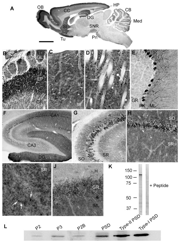
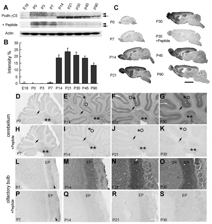
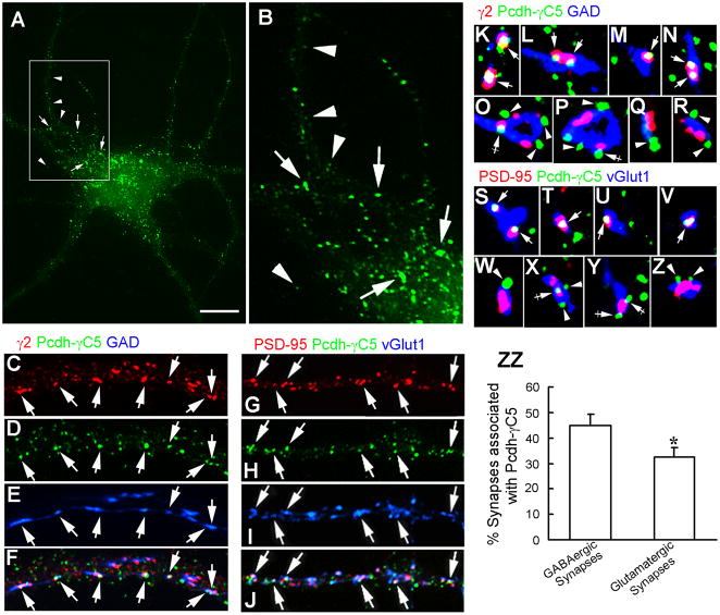
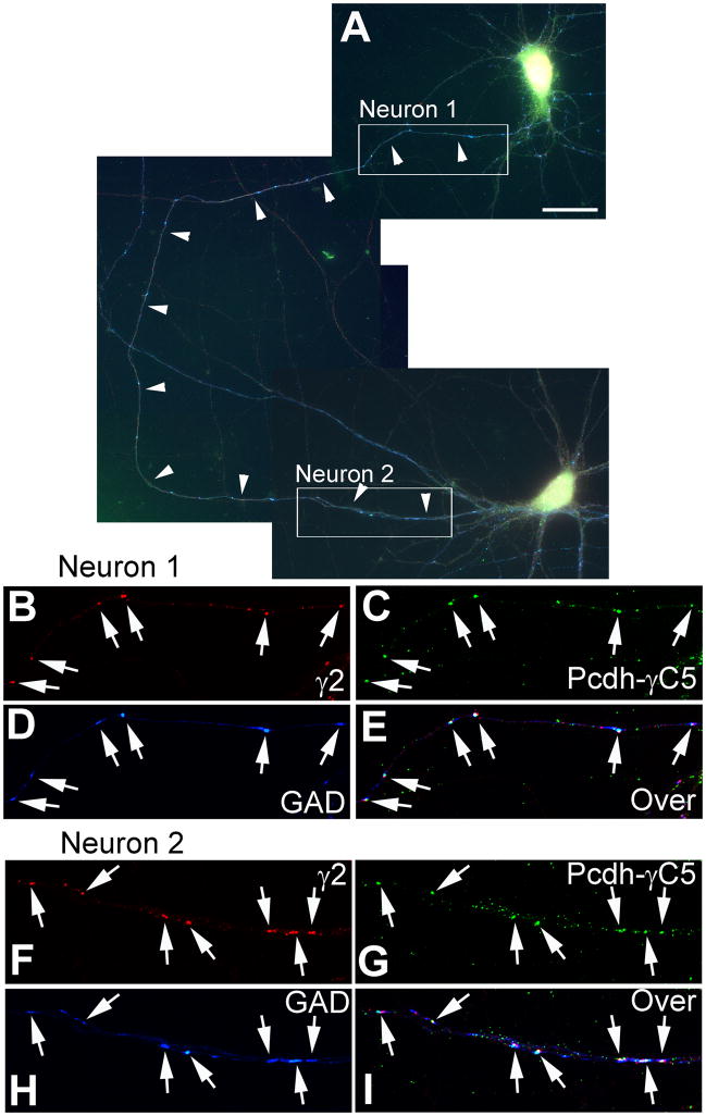
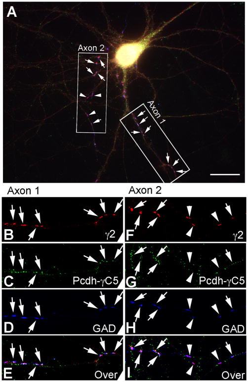
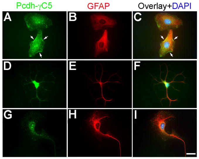
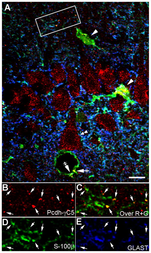
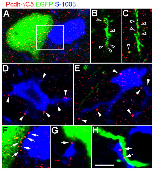
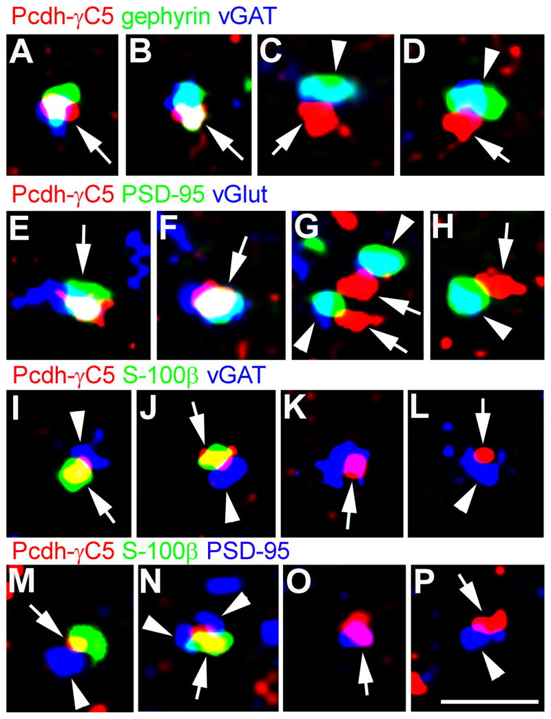
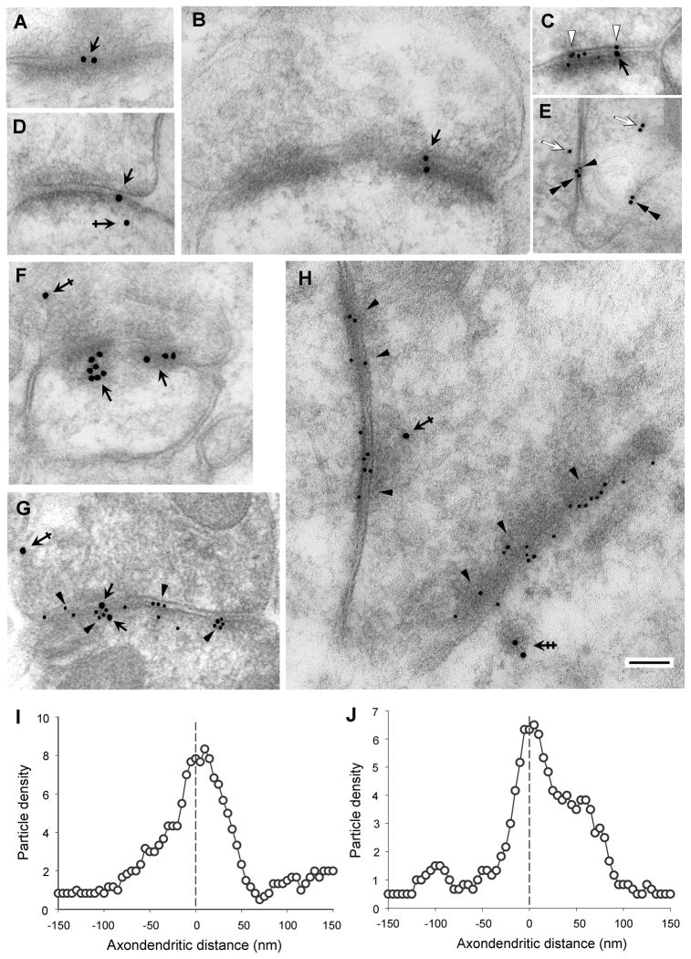
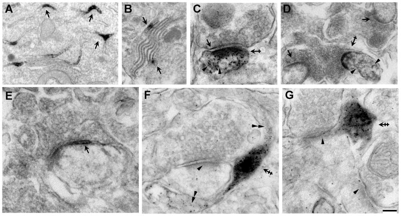
It has been proposed that gamma-protocadherins (Pcdh-gammas) are involved in the establishment of specific patterns of neuronal connectivity. Contrary to the other Pcdh-gammas, which are expressed in the embryo, Pcdh-gammaC5 is expressed postnatally in the brain, coinciding with the peak of synaptogenesis. We have developed an antibody specific for Pcdh-gammaC5 to study the expression and localization of Pcdh-gammaC5 in brain. Pcdh-gammaC5 is highly expressed in the olfactory bulb, corpus striatum, dentate gyrus, CA1 region of the hippocampus, layers I and II of the cerebral cortex, and molecular layer of the cerebellum. Pcdh-gammaC5 is expressed in both neurons and astrocytes. In hippocampal neuronal cultures, and in the absence of astrocytes, a significant percentage of synapses, more GABAergic than glutamatergic, have associated Pcdh-gammaC5 clusters. Some GABAergic axons show Pcdh-gammaC5 in the majority of their synapses. Nevertheless, many Pcdh-gammaC5 clusters are not associated with synapses. In the brain, significant numbers of Pcdh-gammaC5 clusters are located at contact points between neurons and astrocytes. Electron microscopic immunocytochemistry of the rat brain shows that 1) Pcdh-gammaC5 is present in some GABAergic and glutamatergic synapses both pre- and postsynaptically; 2) Pcdh-gammaC5 is also extrasynaptically localized in membranes and in cytoplasmic organelles of neurons and astrocytes; and 3) Pcdh-gammaC5 is also localized in perisynaptic astrocyte processes. The results support the notions that 1) Pcdh-gammaC5 plays a role in synaptic specificity and/or synaptic maturation and 2) Pcdh-gammaC5 is involved in neuron-neuron synaptic interactions and in neuron-astrocyte interactions, including perisynaptic neuron-astrocyte interactions.


Figures

Fig. 1
Fig. 1. Immunocytochemical localization of Pcdh-γC5 in the rat brain and immunoblots of brain fractions
(A) Immunocytochemistry of a parasagittal section of a P30 rat brain with anti-Pcdh-γC5. The Pcdh-γC5 is highly expressed in the olfactory bulb, olfactory tubercle, cerebral cortex, corpus striatum, dentate gyrus, CA1 region of the hippocampus, cerebellum, medial cerebellar nucleus, substantia nigra reticulata and pontine nuclei. (B) Olfactory bulb. (C) Layers I – III of the cerebral cortex. (D) Corpus Striatum. (E) Cerebellum. (F) Hippocampus. (G) The CA2-CA3 region of the hippocampus. (H) The CA1 region of the hippocampus. Note the accumulation of Pcdh-γC5 immunoreactivity in both the neuropil and perikaryon of pyramidal cells. (I) High magnification of the stratum radiatum (SR) of CA1. Note the granular aspect of the immunostaining in the neuropil (arrows). (J) Dentate gyrus and hilus. Scale bar = 3mm in A, 67μm in B, 50μm in C, E and J, 100μm in D, G and H, 250μm in F and 10μm in I. (K) Immunoblot of a rat brain membrane fraction shows that Pcdh-γC5 antibody recognizes a ~120kD protein band that is displaceable by the antigenic peptide (50μg/ml). (L) Immunoblot of various rat brain fractions with Pcdh-γC5 antibody. The 120kD Pcdh-γC5 protein band is present in various brain fractions including the crude synaptosomal (P2), microsomal (P3), synaptosomal (P2B), “One triton” postsynaptic density (PSD), type-II (GABAergic) PSD and type-I (glutamatergic) PSD fractions. The amount of protein loaded in each lane was 4.3μg. The abbreviations used in the panels are: Cerebellum (CB), cerebral cortex (CC), corpus striatum (St), dentate gyrus (DG), external plexiform layer (EP), granule cell layer (GR), glomerular layer (GL), hilus (H), hippocampus (HP), medial cerebellar nucleus (Med), molecular layer (ML), olfactory bulb (OB), olfactory tubercle (Tu), pontine nuclei (Pn), Purkinje cell layer (PK), stratum oriens (SO), stratum pyramidale (SP), stratum radiatum (SR) and substantia nigra reticulata (SNR).
Fig. 2
Fig. 2. Pcdh-γC5 expression during rat brain development
(A) Immunoblots of rat forebrain (telencephalon) homogenates of various ages (42μg of protein per lane). The Pcdh-γC5 antibody recognizes a 120kD protein band (arrow), whose immunoreactivity is displaced by the antigenic peptide (50μg/ml). Between P0 and P7 two proteins of higher Mr (arrowhead) are also recognized by the antibody. However this immunoreactivity is non-specific because the antigenic peptide does not block it and the protein band disappears after P7. An anti-actin antibody showed no significant difference in the protein load in the various lanes. (B) Quantification by densitometry of the Pcdh-γC5 protein band. (C) Light microscopy immunocytochemistry shows the expression of Pcdh-γC5 during the postnatal development of the rat brain. Little or no Pcdh-γC5 is expressed at P0 or P7. At P14 and onwards the brains showed Pcdh-γC5 immunoreactivity, which was blocked by the antigenic peptide (50μg/ml, only shown in this figure for P30). (D–K) Light microscopy immunocytochemistry. Postnatal development of Pcdh-γC5 immunoreactivity in the cerebellum. The single asterisk labels the molecular layer, the circle labels the granule layer, the arrow points to the Purkinje cell layer and the double asterisk labels the deep cerebellar nuclei. (L–S) Light microscopy immunocytochemistry. Postnatal development of Pcdh-γC5 immunoreactivity in the olfactory bulb. For abbreviations in LS, see the legend to Fig. 1. The displacement of the immunoreaction by the antigenic peptide in cerebellum and olfactory bulb is shown in panels HK and PS respectively. Scale bar = 5mm in C; 550μm in DK and 230 μm in LS.
Fig. 3
Fig. 3. Pcdh-γC5 expression in cultured hippocampal neurons
(A) Immunofluorescence of cultured hippocampal neurons (21 DIV) with the Rb anti-Pcdh-γC5 antibody. (B) Enlargement of the inset box in A. Arrows point to larger Pcdh-γC5 normally found in the soma and proximal dendrites. Arrowheads point to smaller clusters that are present in dendrites. (C–F) Triple-label immunofluorescence with GP anti-γ2 (C), Rb anti-Pcdh-γC5 (D), sheep anti-GAD (E) and overlay (F). (G–J) Triple label immunofluorescence with mouse anti-PSD95 (G), Rb anti-Pcdh-γC5 (H), GP anti-vGluT1 (I) and overlay (J). Arrows show GABAergic (CF) and glutamatergic (GJ) synapses that have colocalizing Pcdh-γC5 clusters. (K–R) High magnification images show the association of Pcdh-γC5 with GABAergic synapses. Pcdh-γC5 clusters colocalize with synaptic γ2 clusters (KN, arrows) or partially colocalize with synaptic γ2 clusters (O and P, crossed arrows) or partially or fully colocalize with GAD terminals but not with γ2 (OR, arrowheads). (S–Z) Association of Pcdh-γC5 with glutamatergic synapses. Pcdh-γC5 clusters colocalize with synaptic PSD-95 (SV, arrows) or partially colocalize with synaptic PSD-95 clusters (X and Y, crossed arrows) or partially or fully colocalize with vGlut1 terminals but not PSD-95 (W, X, Z, arrowheads). (ZZ) Quantification of the % of GABAergic and glutamatergic synapses that show associated Pcdh-γC5 clusters. Scale bar = 13μm for A, 5μm for BJ and 1.5μm for KZ. * p<0.05 in Student’s t test.
Fig. 4
Fig. 4. Some GABAergic axons have Pcdh-γC5 in the majority of their synapses
(A) Overlay of triple-label immunofluorescence of 21 DIV cultured hippocampal neurons showing the same axon (arrowheads) contacting two different pyramidal cells (neuron 1 and neuron 2). (B–I) Enlargements of the inset boxes in A shows GABAergic synapses from the same axon on neuron 1 (BE) and neuron 2 (FI). Triple-label immunofluorescence with Rb anti-Pcdh-γC5 (green), GP anti-γ2 (red) and sheep anti-GAD (blue) shows that the majority of the GABAergic synapses (GAD+ and γ2+, arrows) from this axon on the two neurons have colocalizing Pcdh-γC5 clusters. Scale bar = 24 μm for A and 10μm for BI.
Fig. 5
Fig. 5. Two GABAergic axons contacting the same neuron show differential extent of Pcdh-γC5 localization to their synapses
(A) Overlay of triple-label immunofluorescence of 21 DIV cultured hippocampal neurons showing two axons contacting a pyramidal cell (axon 1 and axon 2). (B–I) Enlargements of the insets in A shows GABAergic synapses (arrows) from axon 1 (BE) and axon 2 (FI). Triple-label immunofluorescence with Rb anti-Pcdh-γC5 (green), GP anti-γ2 (red) and sheep anti-GAD (blue). The majority of the GABAergic synapses (GAD+ and γ2+, arrows) from axon 1 (BE) have colocalizing Pcdh-γC5 clusters. In contrast, axon 2 has some GABAergic synapses with Pcdh-γC5 (FI, arrows) and some without Pcdh-γC5 (FI, arrowheads). Scale bar = 20μm for A and 13μm for BI.
Fig. 6
Fig. 6. Pcdh-γC5 is expressed by cultured astrocytes
Triple-label fluorescence of cultured astrocytes with Rb anti-Pcdh-γC5 (green, A, D, G), mouse anti-GFAP (red, B, E, H) and the nuclear stain DAPI (blue, C, F, I). The Pcdh-γC5 is expressed in astrocytes with various morphologies. The Pcdh-γC5 immunofluorescence is frequently shown in the form of clusters both in the cell body and processes. Some of the clusters are clearly associated with the plasma membrane (A, C, arrows). Scale bar=20μm.
Fig. 7
Fig. 7. In the intact brain, Pcdh-γC5 clusters are present in neurons and astrocytes
Triple-label laser confocal microscopy of the CA1 region of the hippocampus in the P83 rat brain with Rb anti-Pcdh-γC5 (red), mouse anti-S-100β (green) and GP anti-GLAST (blue). (A) Pcdh-γC5 clusters are present in the soma of neurons (double arrowheads), cell bodies of astrocytes (arrowheads) and the astrocytic end-feet contacting blood vessels (crossed arrows). (B–E) The high magnification image of the inset box in panel A shows that Pcdh-γC5 clusters are present in astrocytic processes (arrows) revealed with antibodies to S-100β and GLAST. The scale bar = 10 μm in A and 6μm in BE.
Fig. 8
Fig. 8. In the intact brain Pcdh-γC5 forms clusters at contact point between neurons and astrocytes
(A–H) Laser confocal microscopy of the cerebral cortex of a P35 rat brain in which a limited population of neurons are labeled by EGFP (green) by in utero electroporation. Single optical section 0.1μm thick. The tissue was triple-label with EGFP fluorescence (green) and antibodies to Pcdh-γC5 (red) and S-100β (blue). Pcdh-γC5 forms clusters on the neuronal soma (A) and dendrites (empty arrowheads in B and C), astrocyte cell body and processes (D and E), at contact points between neuron and astrocyte (arrows in F, G and H). Note the presence Pcdh-γC5 clusters at the edge of astrocytes (arrowheads in D and E). The scale bar = 5 μm in panels A to E and 2.8 μm in panels F to H.
Fig. 9
Fig. 9. Synaptic and perisynaptic Pcdh-γC5 clusters in the intact brain
Laser confocal microscopy of the CA1 region of the hippocampus. (A–B) Some Pcdh-γC5 clusters colocalize (arrows) with the GABAergic synaptic markers gephyrin and vGAT. (C–D) Some Pcdh-γC5 clusters (arrows) are adjacent to the GABAergic synaptic markers vGAT and gephyrin (arrowheads). (E–F) Some Pcdh-γC5 clusters colocalize (arrows) with the glutamatergic synaptic markers PSD-95 and vGlut1. (G–H) Some Pcdh-γC5 clusters (arrows) are adjacent to the glutamatergic synaptic markers PSD-95 and vGlut1 (arrowheads). (I–J) Some Pcdh-γC5 clusters (arrows) are on S-100β+ astrocytic processes adjacent to vGAT puncta (arrowheads). (K–L) Some Pcdh-γC5 clusters (arrows) that colocalize (K) or are adjacent to vGAT puncta (arrowhead, L) are not associated with the astrocytic marker S-100β. (M–N) Some Pcdh-γC5 clusters (arrows) adjacent to the glutamatergic synaptic marker PSD-95 (arrowheads) are on S-100β+ astrocytic processes. (O–P) Some Pcdh-γC5 clusters (arrows) that colocalize (O) or are adjacent to PSD-95 clusters (arrowhead, P) are not associated with the astrocyte marker S-100β. Scale bar = 1μm.
Fig. 10
Fig. 10. Post-embedding EM immunogold of rat brain with anti-Pcdh-γC5 antibody
(A–H) Post-embedding EM immunogold labeling with Rb Pcdh-γC5 in single-label experiment (A, B, D and F) or in double-label experiment with mouse mAb anti-GluR2 (C) or mouse mAb anti-β2/3 GABAAR (E, G and H). In all panels except E, where there is no synapse, the presynaptic terminal is located above the synaptic cleft. The larger gold particles (18 nm diameter) correspond to Pcdh-γC5 immunoreactivity, while the small particles (10 nm) correspond to GluR2 immunoreactivity in C or to β2/3 GABAAR immunoreactivity in E, G and H. The Pcdh-γC5 immunoreactivity is associated with the synaptic complex (A,B,C,D,F, and G, black arrows), presynaptic terminals and postsynaptic cytoplasm (D, F, G and H, single and double crossed arrows) and non-synaptic membranes (E, double arrowheads) and cytoplasm (E, white arrows). The double-crossed arrow in H points to Pcdh-γC5 immunoreactivity associated with a postsynaptic vesicular structure. Single black arrowheads point to GABAAR immunoreactivity (small particles) in GABAergic synapses, while in E the single black arrowhead points to a GABAAR gold particle associated with a non-synaptic membrane. White arrowheads in C point to GluR2 immunoreactivity. Thin sections are from the cerebral cortex (A,B,D, E and F) or from the molecular layer of the cerebellum (C, G and H). The scale bar represents 100 nm in all panels except in panel E (160nm). (I and J) Axodendritic distribution of Pcdh-γC5 gold particles (n=167) in GABAergic synapses (I) or in glutamatergic/type I synapses (J, n=133). Gold particles are distributed in 5 nm bins according to the distance from the center of each gold particle to the midline of the synaptic cleft defined as the zero point. Negative and positive values represent presynaptic and postsynaptic localization respectively. Particle density is the number of particles in a 5-nm bin. The graph was smoothed using a six-point weighted running average.
Fig. 11
Fig. 11. Pre-embedding EM immunocytochemistry of the rat brain with anti-Pcdh-γC5 antibody
(A) Immunoreactivity with the Pcdh-γC5 antibody concentrates in the endoplasmic reticulum of some neurons (arrows). (B) Immunoreactivity in Purkinje cell cisternae (arrows). (C and D) Some synapses with type-I morphology show immunoreactivity in synaptic membranes (C, arrow) and particularly in the postsynaptic density (C and D, crossed arrows). Immunoreactivity was also found in the postsynaptic cytoplasm separated from the postsynaptic density (C and D, arrowheads). Note in D that one synapse shows immunoreactivity (crossed arrows) while two others show no immunoreactivity (arrows). (E) Some synapses with type-II morphology show immunoreactivity (arrow) in the pre- and post-synaptic membranes and in the synaptic cleft. (F and G) Some glial processes (double crossed arrows) adjacent to unlabeled synapses (single arrowheads) show strong immunoreactivity. Little or no accumulation of immunoreactivity is found in the same glial processes distant from the synapses (double arrowheads). Samples are from hippocampus (A), cerebellum (B, D and E) and cerebral cortex (C, F and G). The brain was fixed wit either 4% paraformaldehyde, 0.1% glutaraldehyde (A, C, F and G) or with PLP (B, D and E). The scale bar represents 100 nm for C, E, F and G; 162 nm for D; 270 nm for B; and 325 nm for A.

Similar articles

Cited by

References

    1. Altman J. Postnatal development of the cerebellar cortex in the rat. II. Phases in the maturation of Purkinje cells and of the molecular layer. J Comp Neurol. 1972;145:399–463. - PubMed
    1. Blank M, Triana-Baltzer GB, Richards CS, Berg DK. Alpha-protocadherins are presynaptic and axonal in nicotinic pathways. Mol Cell Neurosci. 2004;26:530–543. - PubMed
    1. Bonn S, Seeburg PH, Schwarz MK. Combinatorial expression of alpha- and gamma-protocadherins alters their presenilin-dependent processing. Mol Cell Biol. 2007;27:4121–4132. - PMC - PubMed
    1. Charych EI, Yu W, Li R, Serwanski DR, Miralles CP, Li X, Yang BY, Pinal N, Walikonis R, De Blas AL. A four PDZ domain-containing splice variant form of GRIP1 is localized in GABAergic and glutamatergic synapses in the brain. J Biol Chem. 2004a;279:38978–38990. - PubMed
    1. Charych EI, Yu W, Miralles CP, Serwanski DR, Li X, Rubio M, De Blas AL. The brefeldin A-inhibited GDP/GTP exchange factor 2, a protein involved in vesicular trafficking, interacts with the beta subunits of the GABA receptors. J Neurochem. 2004b;90:173–189. - PubMed

Publication types

MeSH terms