

Identification of disulfide bonds in protein proteolytic degradation products using de novo-protein unique sequence tags approach
- PMID: 20590115
- PMCID: PMC3217045
- DOI: 10.1021/pr1002559
Identification of disulfide bonds in protein proteolytic degradation products using de novo-protein unique sequence tags approach
Abstract
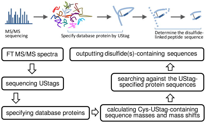
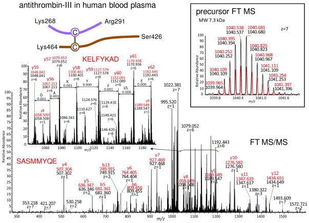
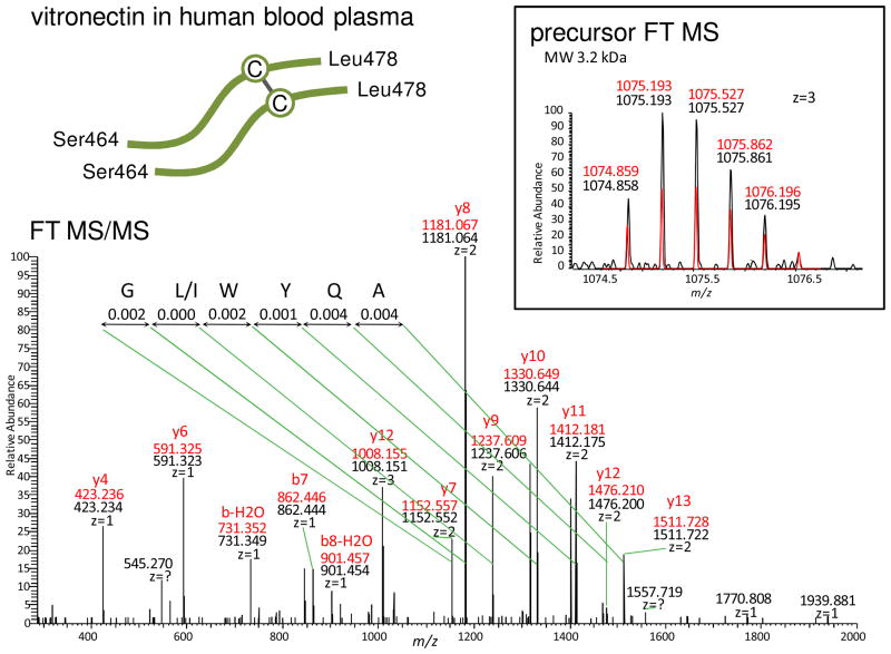
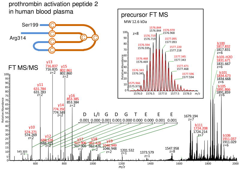
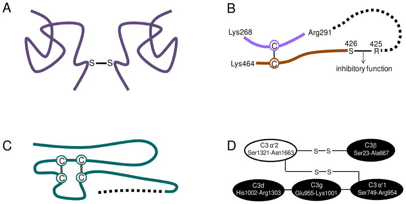
Disulfide bonds are a form of post-translational modification that often determines protein structure(s) and function(s). In this work, we report a mass spectrometry method for identification of disulfides in degradation products of proteins, specifically endogenous peptides in the human blood plasma peptidome. LC-Fourier transform tandem mass spectrometry (FT MS/MS) was used for acquiring mass spectra that were de novo sequenced and then searched against the IPI human protein database. Through the use of unique sequence tags (UStags), we unambiguously correlated the spectra to specific database proteins. Examination of the UStags' prefix and/or suffix sequences that contain cysteine(s) in conjunction with sequences of the UStags-specified database proteins is shown to enable the unambigious determination of disulfide bonds. Using this method, we identified the intermolecular and intramolecular disulfides in human blood plasma peptidome peptides that have molecular weights of up to approximately 10 kDa.
Figures
Similar articles
-
De novo sequencing of unique sequence tags for discovery of post-translational modifications of proteins.Anal Chem. 2008 Oct 15;80(20):7742-54. doi: 10.1021/ac801123p. Epub 2008 Sep 11. Anal Chem. 2008. PMID: 18783246 Free PMC article.
-
Characterization of disulfide bonds by planned digestion and tandem mass spectrometry.Mol Biosyst. 2015 Apr;11(4):1156-64. doi: 10.1039/c4mb00688g. Mol Biosyst. 2015. PMID: 25703060 Free PMC article.
-
Generic Workflow for Mapping of Complex Disulfide Bonds Using In-Source Reduction and Extracted Ion Chromatograms from Data-Dependent Mass Spectrometry.Anal Chem. 2018 Jul 3;90(13):8202-8210. doi: 10.1021/acs.analchem.8b01603. Epub 2018 Jun 22. Anal Chem. 2018. PMID: 29878755
-
The spectral networks paradigm in high throughput mass spectrometry.Mol Biosyst. 2012 Oct;8(10):2535-44. doi: 10.1039/c2mb25085c. Mol Biosyst. 2012. PMID: 22610447 Free PMC article. Review.
-
Software eyes for protein post-translational modifications.Mass Spectrom Rev. 2015 Mar-Apr;34(2):133-47. doi: 10.1002/mas.21425. Epub 2014 Jun 2. Mass Spectrom Rev. 2015. PMID: 24889695 Review.
Cited by
-
Facilitating protein disulfide mapping by a combination of pepsin digestion, electron transfer higher energy dissociation (EThcD), and a dedicated search algorithm SlinkS.Mol Cell Proteomics. 2014 Oct;13(10):2776-86. doi: 10.1074/mcp.O114.039057. Epub 2014 Jun 30. Mol Cell Proteomics. 2014. PMID: 24980484 Free PMC article.
-
Blood peptidome-degradome profile of breast cancer.PLoS One. 2010 Oct 18;5(10):e13133. doi: 10.1371/journal.pone.0013133. PLoS One. 2010. PMID: 20976186 Free PMC article.
-
Effectiveness of CID, HCD, and ETD with FT MS/MS for degradomic-peptidomic analysis: comparison of peptide identification methods.J Proteome Res. 2011 Sep 2;10(9):3929-43. doi: 10.1021/pr200052c. Epub 2011 Aug 15. J Proteome Res. 2011. PMID: 21678914 Free PMC article.
References
-
- Christis C, Lubsen NH, Braakman I. Protein folding includes oligomerization – examples from the endoplasmic reticulum and cytosol. FEBS J. 2008;275:4700–4727. - PubMed
-
- Riemer J, Bulleid N, Herrmann JM. Disulfide formation in the ER and mitochondria: two solutions to a common process. Science. 2009;324:1284–1287. - PubMed
-
- Dranoff G. Targets of protective tumor immunity. Ann N Y Acad Sci. 2009;1174:74–80. - PubMed
-
- Hogg PJ. Contribution of allosteric disulfide bonds to regulation of hemostasis. J Thromb Haemost. 2009;7:12–16. - PubMed
-
- Nakamura T, Lipton SA. Cell death: protein misfolding and neurodegenerative diseases. Apoptosis. 2009;14:455–468. - PubMed
Publication types
MeSH terms
Substances
Grants and funding
LinkOut - more resources
Full Text Sources
