Reflex conditioning: a new strategy for improving motor function after spinal cord injury
- PMID: 20590534
- PMCID: PMC2925434
- DOI: 10.1111/j.1749-6632.2010.05565.x
Reflex conditioning: a new strategy for improving motor function after spinal cord injury
Abstract
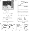
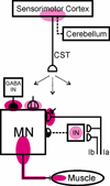
Spinal reflex conditioning changes reflex size, induces spinal cord plasticity, and modifies locomotion. Appropriate reflex conditioning can improve walking in rats after spinal cord injury (SCI). Reflex conditioning offers a new therapeutic strategy for restoring function in people with SCI. This approach can address the specific deficits of individuals with SCI by targeting specific reflex pathways for increased or decreased responsiveness. In addition, once clinically significant regeneration can be achieved, reflex conditioning could provide a means of reeducating the newly (and probably imperfectly) reconnected spinal cord.
Figures


Similar articles
-
Operant conditioning of a spinal reflex can improve locomotion after spinal cord injury in humans.J Neurosci. 2013 Feb 6;33(6):2365-75. doi: 10.1523/JNEUROSCI.3968-12.2013. J Neurosci. 2013. PMID: 23392666 Free PMC article.
-
Could enhanced reflex function contribute to improving locomotion after spinal cord repair?J Physiol. 2001 May 15;533(Pt 1):75-81. doi: 10.1111/j.1469-7793.2001.0075b.x. J Physiol. 2001. PMID: 11351015 Free PMC article. Review.
-
H-reflex conditioning during locomotion in people with spinal cord injury.J Physiol. 2021 May;599(9):2453-2469. doi: 10.1113/JP278173. Epub 2019 Jul 11. J Physiol. 2021. PMID: 31215646 Free PMC article.
-
Restoring walking after spinal cord injury: operant conditioning of spinal reflexes can help.Neuroscientist. 2015 Apr;21(2):203-15. doi: 10.1177/1073858414527541. Epub 2014 Mar 17. Neuroscientist. 2015. PMID: 24636954 Free PMC article. Review.
-
Persistent beneficial impact of H-reflex conditioning in spinal cord-injured rats.J Neurophysiol. 2014 Nov 15;112(10):2374-81. doi: 10.1152/jn.00422.2014. Epub 2014 Aug 20. J Neurophysiol. 2014. PMID: 25143542 Free PMC article.
Cited by
-
Neural adaptations to electrical stimulation strength training.Eur J Appl Physiol. 2011 Oct;111(10):2439-49. doi: 10.1007/s00421-011-2012-2. Epub 2011 Jun 4. Eur J Appl Physiol. 2011. PMID: 21643920 Free PMC article. Review.
-
The effects of anodal transcranial direct current stimulation and patterned electrical stimulation on spinal inhibitory interneurons and motor function in patients with spinal cord injury.Exp Brain Res. 2016 Jun;234(6):1469-78. doi: 10.1007/s00221-016-4561-4. Epub 2016 Jan 20. Exp Brain Res. 2016. PMID: 26790423 Free PMC article.
-
Electrical Stimulation of Low-Threshold Proprioceptive Fibers in the Adult Rat Increases Density of Glutamatergic and Cholinergic Terminals on Ankle Extensor α-Motoneurons.PLoS One. 2016 Aug 23;11(8):e0161614. doi: 10.1371/journal.pone.0161614. eCollection 2016. PLoS One. 2016. PMID: 27552219 Free PMC article.
-
Changes in H-reflex and V-waves following spinal manipulation.Exp Brain Res. 2015 Apr;233(4):1165-73. doi: 10.1007/s00221-014-4193-5. Epub 2015 Jan 13. Exp Brain Res. 2015. PMID: 25579661
-
Biological basis of exercise-based treatments: spinal cord injury.PM R. 2011 Jun;3(6 Suppl 1):S73-7. doi: 10.1016/j.pmrj.2011.02.019. PM R. 2011. PMID: 21703584 Free PMC article. Review.
References
-
- Matthews PBC. Mammalian Muscle Receptors and their Central Actions. Baltimore, MD: Williams & Wilkins; 1972. pp. 319–409.
-
- Lee RG, Tatton WG. Motor responses to sudden limb displacement in primate with specific CNS lesions and in human patients with motor system disorders. Can. J. Neurol. Sci. 1975;2:285–293. - PubMed
-
- Burke RE, Rudomin P. Spinal neurons and synapses. In: Brooks VB, editor. Handbook of Physiology. Section I: The Nervous System. Volume I: Cellular Biology of Neurons. Part II. Baltimore, MD: Williams and Wilkins Company; 1977. pp. 877–944.
-
- Baldissera F, Hultborn H, Illert M. Integration in Spinal Neuronal Systems. In: Brooks VB, editor. Handbook of Physiology. Section I: The Nervous System. Volume II: Motor Control, Part I. Baltimore, MD: Williams and Wilkins Company; 1981. pp. 509–595.
-
- Henneman E, Mendell LM. Functional organization of motoneuron pool and inputs. In: Brooks VB, editor. Handbook of Physiology. Section I: The Nervous System. Volume II: Motor Control, Part I. Baltimore, MD: Williams and Wilkins Company; 1981. pp. 423–507.
Publication types
MeSH terms
Grants and funding
LinkOut - more resources
Full Text Sources
Other Literature Sources
Medical

