Review
. 2010 Jun;160(3):423-42.
doi: 10.1111/j.1476-5381.2010.00787.x.

Quantification of brain endocannabinoid levels: methods, interpretations and pitfalls

Affiliations
Review

Quantification of brain endocannabinoid levels: methods, interpretations and pitfalls

Matthew W Buczynski et al. Br J Pharmacol. 2010 Jun.

Abstract

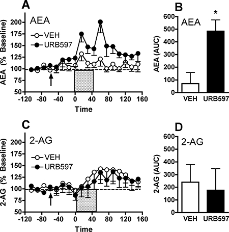
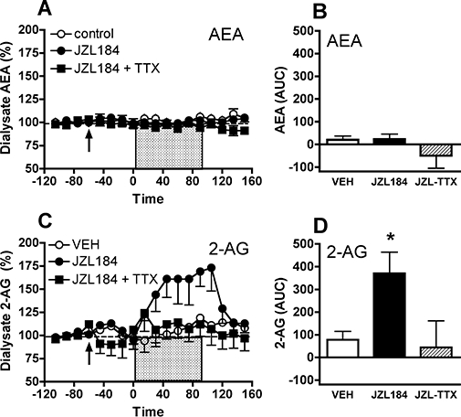
Endocannabinoids play an important role in a diverse range of neurophysiological processes including neural development, neuroimmune function, synaptic plasticity, pain, reward and affective state. This breadth of influence and evidence for altered endocannabinoid signalling in a variety of neuropathologies has fuelled interest in the accurate quantification of these lipids in brain tissue. Established methods for endocannabinoid quantification primarily employ solvent-based lipid extraction with further sample purification by solid phase extraction. In recent years in vivo microdialysis methods have also been developed for endocannabinoid sampling from the brain interstitial space. However, considerable variability in estimates of endocannabinoid content has led to debate regarding the physiological range of concentrations present in various brain regions. This paper provides a critical review of factors that influence the quantification of brain endocannabinoid content as determined by lipid extraction from bulk tissue and by in vivo microdialysis. A variety of methodological issues are discussed including analytical approaches, endocannabinoid extraction and purification, post-mortem changes in brain endocannabinoid content, cellular reactions to microdialysis probe implantation and caveats related to lipid sampling from the extracellular space. The application of these methods for estimating brain endocannabinoid content and the effects of endocannabinoid clearance inhibition are discussed. The benefits, limitations and pitfalls associated with each approach are emphasized, with an eye toward the appropriate interpretation of data gathered by each method.


Figures

Figure 1
Figure 1
Comparison of published estimates of brain tissue endocannabinoid content in six brain regions of drug-naïve rats. Data are compiled from 30 publications in which both AEA (anandamide, N-arachidonoyl-ethanolamine) and 2-arachidonoylglycerol (2-AG) were quantified in tissue samples from multiple brain regions, as well as whole brain for comparison. Each small data point represents the average value of AEA and 2-AG content reported in an individual study, with average values from at least eight publications presented for each brain region. The mean and standard error of the collective AEA and 2-AG measures are represented as large ovals.
Figure 2
Figure 2
Effect of perfusate hydroxy-propyl-β-cyclodextrin (HP-β-CD) content on dialysate endocannabinoid (eCB) levels. (A) Evaluation of in vitro eCB sampling efficiency. Dialysis probes (n= 6) were suspended in a medium containing 200 nM AEA (anandamide, N-arachidonoyl-ethanolamine) and 2-arachidonoylglycerol (2-AG), and dialysates were collected with aCSF perfusates containing 0–30% HP-β-CD. (B) Effect of perfusate HP-β-CD (0–30%) on eCB content in dialysates collected from the rat nucleus accumbens (n= 6). In both experiments dialysate AEA and 2-AG levels were below limits of quantification (n.d.) in samples collected without perfusate HP-β-CD. Inclusion of 10% HP-β-CD in the perfusate significantly enhanced dialysate eCB recovery, and higher HP-β-CD perfusate levels did not further improve dialysate eCB content collected either in vitro or in vivo.
Figure 3
Figure 3
Effect of euthanasia on in vivo microdialysate endocannabinoid levels. Microdialysis samples were collected from the nucleus accumbens of naïve Wistar rats (n= 6) at 10 min intervals starting 90 min prior to euthanasia by CO2 narcosis (induced at time zero) until 60 min post-mortem. (A) Dialysate 2-arachidonoylglycerol (2-AG) levels were stable over the 90 min pre-euthanasia baseline period (repeated measures anova, F5,45= 0.578, not significant), but were significantly increased during the 60 min post-mortem sampling period (F5,30= 32.635, P < 0.0001). (B) AEA (anandamide, N-arachidonoyl-ethanolamine) levels measured in these same samples were also stable during the baseline period (F5,45= 1.824, not significant) and were not significantly altered during the 60 min post-mortem period (F5,30= 1.367, not significant). The double asterisks denote P < 0.01 versus pre-euthanasia baseline at individual post-mortem time points as determined by Fisher's post hoc comparisons following anova. See text for additional details.
Figure 4
Figure 4
Effect of systemic URB597 administration on AEA (anandamide, N-arachidonoyl-ethanolamine) and 2-arachidonoylglycerol (2-AG) levels in microdialysates collected from rat nucleus accumbens. Baseline dialysate AEA levels were 1.7 ± 0.6 nM in animals in the URB597 group (n= 7) and 1.9 ± 0.8 nM in animals in the vehicle (VEH) group (n= 6). Baseline dialysate 2-AG levels were 10.2 ± 1.0 and 9.4 ± 1.2 nM in these groups respectively. (A) Administration of 0.3 mg·kg−1 URB597 (arrow, time 0 min) induced a significant increase in dialysate AEA levels observable 60 min after administration that persisted for over 2 h. (B) Group comparison of URB597 effect as determined by the area under the curve (AUC). (C and D) Levels of 2-AG measured in these same dialysate samples were not significantly altered by either URB597 or vehicle administration. The asterisk denotes P < 0.05 between URB and VEH groups, as determined by Fisher's post hoc evaluations following anova (A and C) or by anova of group AUC values (B and D).
Figure 5
Figure 5
Effect of the fatty acid amide hydrolase inhibitor URB597 on depolarization-induced increases in dialysate AEA (anandamide, N-arachidonoyl-ethanolamine) and 2-arachidonoylglycerol (2-AG) levels in the rat nucleus accumbens. Baseline dialysate AEA levels were 1.9 ± 0.4 nM in animals in the URB597 group (n= 7) and 2.1 ± 0.9 nM in animals in the vehicle (VEH) group (n= 6). Baseline dialysate 2-AG levels were 9.4 ± 1.1 and 8.3 ± 1.5 nM respectively. URB597 (0.3 mg·kg−1, i.p.) or saline were administered 60 min prior to delivery of high KCl (90 mM) and CaCl2 (10 mM) perfusate for 45 min. (A) High K+/Ca2+ solution (shaded bar) induced a subtle, transient increase in dialysate AEA that was more pronounced in rats pretreated with URB597. (B) Group comparison of URB597 effect as determined by the area under the curve (AUC). (C and D) High K+/Ca2+ solution induced a similar transient change in dialysate 2-AG levels; URB597 pretreatment had no effect on this profile. The asterisk denotes P < 0.05 between URB and VEH groups, as determined by Fisher's post hoc evaluations following anova (A and C) or by anova of group AUC values (B and D).
Figure 6
Figure 6
Effect of the monoacylglycerol lipase inhibitor JZL184 on dialysate AEA (anandamide, N-arachidonoyl-ethanolamine) and 2-arachidonoylglycerol (2-AG) levels in the nucleus accumbens of C57BL/6 mice. JZL184 (10 mg·kg−1, i.p.) or saline were administered 60 min prior to a 90 min perfusion of high concentrations of KCl (150 mM) and CaCl2 (10 mM). High K+/Ca2+ solution (shaded bar) did not significantly alter dialysate AEA levels, as determined (A) by temporal analysis or (B) by area under the curve (AUC) comparison of the experimental groups. Baseline dialysate AEA levels were 0.54 ± 0.1 nM for the JZL184 group (n= 8), 0.55 ± 0.1 nM for the JZL184 + TTX (tetrodotoxin) group (n= 6) and 0.58 ± 0.08 nM for the vehicle (VEH) group (n= 6). (C and D) Depolarization subtly increased dialysate 2-AG levels in vehicle-treated mice, and JZL184 pretreatment substantially enhanced this effect. Co-perfusion with TTX blocked the effects of high K+/Ca2+ and JZL184, confirming that dialysate 2-AG increases in an impulse-dependent manner. The asterisk denotes P < 0.05 between JZL and VEH groups, as determined by Fisher's post hoc evaluations following anova (A and C) or by anova of group AUC values (B and D). Baseline dialysate 2-AG levels were 4.6 ± 0.7 nM for the JZL184 group, 4.4 ± 0.3 nM for the JZL184 + TTX group and 4.2 ± 0.4 nM for the vehicle group.

References

    1. Abulrob A, Tauskela JS, Mealing G, Brunette E, Faid K, Stanimirovic D. Protection by cholesterol-extracting cyclodextrins: a role for N-methyl-D-aspartate receptor redistribution. J Neurochem. 2005;92:1477–1486. - PubMed
    1. Ahn K, Johnson DS, Mileni M, Beidler D, Long JZ, McKinney MK, et al. Discovery and characterization of a highly selective FAAH inhibitor that reduces inflammatory pain. Chem Biol. 2009;16:411–420. - PMC - PubMed
    1. Alexander SP, Mathie A, Peters JA. Guide to Receptors and Channels (GRAC), 3rd edition. Br J Pharmacol. 2008;153(Suppl. 2):S1–209. - PMC - PubMed
    1. Alger BE. Retrograde signaling in the regulation of synaptic transmission: focus on endocannabinoids. Prog Neurobiol. 2002;68:247–286. - PubMed
    1. Alvarez-Jaimes L, Polis I, Parsons LH. Regional influence of CB1 receptor signaling on ethanol self-administration by rats. The Open Neuropsychopharmacology. 2009a;2:77–85. - PMC - PubMed

Publication types

MeSH terms

Substances