. 2010 Jun;160(3):643-56.
doi: 10.1111/j.1476-5381.2010.00710.x.

Regulation of Fas receptor/Fas-associated protein with death domain apoptotic complex and associated signalling systems by cannabinoid receptors in the mouse brain

Affiliations

Regulation of Fas receptor/Fas-associated protein with death domain apoptotic complex and associated signalling systems by cannabinoid receptors in the mouse brain

M Alvaro-Bartolomé et al. Br J Pharmacol. 2010 Jun.

Abstract

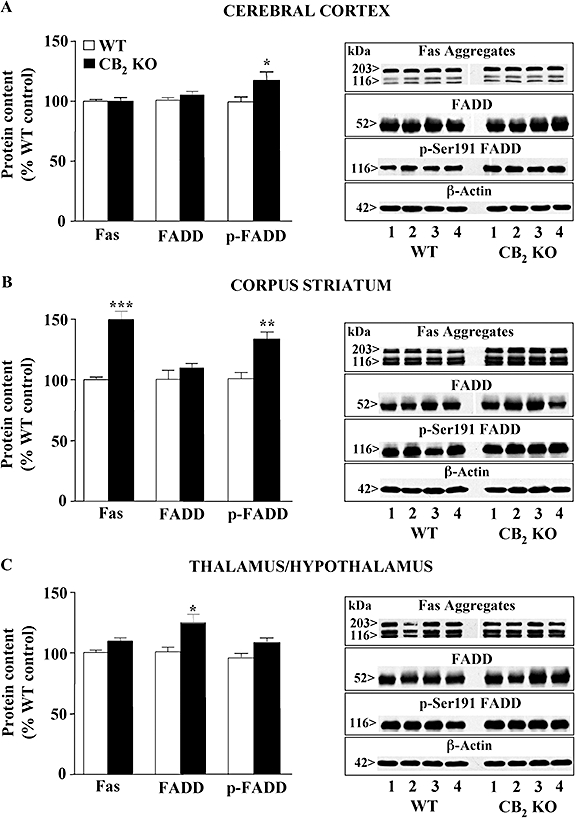
Background and purpose: Natural and synthetic cannabinoids (CBs) induce deleterious or beneficial actions on neuronal survival. The Fas-associated protein with death domain (FADD) promotes apoptosis, and its phosphorylated form (p-FADD) mediates non-apoptotic actions. The regulation of Fas/FADD, mitochondrial apoptotic proteins and other pathways by CB receptors was investigated in the mouse brain.

Experimental approach: Wild-type, CB(1) and CB(2) receptor knock-out (KO) mice were used to assess differences in receptor genotypes. CD1 mice were used to evaluate the effects of CB drugs on canonical apoptotic pathways and associated signalling systems. Target proteins were quantified by Western blot analysis.

Key results: In brain regions of CB(1) receptor KO mice, Fas/FADD was reduced, but p-Ser191 FADD and the p-FADD/FADD ratio were increased. In CB(2) receptor KO mice, Fas/FADD was increased, but the p-FADD/FADD ratio was not modified. In mutant mice, cleavage of poly(ADP-ribose)-polymerase (PARP) did not indicate alterations in brain cell death. In CD1 mice, acute WIN55212-2 (CB(1) receptor agonist), but not JWH133 (CB(2) receptor agonist), inversely modulated brain FADD and p-FADD. Chronic WIN55212-2 induced FADD down-regulation and p-FADD up-regulation. Acute and chronic WIN55212-2 did not alter mitochondrial proteins or PARP cleavage. Acute, but not chronic, WIN55212-2 stimulated activation of anti-apoptotic (ERK, Akt) and pro-apoptotic (JNK, p38 kinase) pathways.

Conclusions and implications: CB(1) receptors appear to exert a modest tonic activation of Fas/FADD complexes in brain. However, chronic CB(1) receptor stimulation decreased pro-apoptotic FADD and increased non-apoptotic p-FADD. The multifunctional protein FADD could participate in the mechanisms of neuroprotection induced by CBs.


Figures

Figure 1
Figure 1
Effects of constitutive deletion of CB1 receptors (WT, n= 9; CB1 KO, n= 9) on the basal contents of Fas receptor (∼116/203 kDa aggregated forms), FADD (∼52 kDa dimeric form) and p-Ser191 FADD (∼116 kDa oligomeric form) in the mouse cerebral cortex (A), corpus striatum (B) and cerebellum (C). The columns are means ± SEM values (% immunoreactivity) expressed as percentage of WT control mice. *P < 0.05; **P < 0.01 when compared with the corresponding WT group (two-tailed Student's t-test). Right: representative immunoblots for Fas, FADD, p-FADD and β-actin in the various brain regions of WT (n= 4) and CB1 KO (n= 4) mice. Protein molecular masses (kDa) were estimated from referenced standards.
Figure 2
Figure 2
Effects of constitutive deletion of CB2 receptors (WT, n= 12; CB2 KO, n= 13) on the basal contents of Fas receptor (∼116/203 kDa aggregated forms), FADD (∼52 kDa dimeric form) and p-Ser191 FADD (∼116 kDa oligomeric form) in the mouse cerebral cortex (A), corpus striatum (B) and thalamus/hypothalamus (C). The columns are means ± SEM values (% immunoreactivity), expressed as percentage of WT control mice. *P < 0.05; **P < 0.01; ***P < 0.0001 when compared with the corresponding WT group (Student's t-test). Right: representative immunoblots for Fas, FADD, p-FADD and β-actin in the various brain regions of WT (n= 4) and CB2 KO (n= 4) mice. Protein molecular masses (kDa) were estimated from referenced standards.
Figure 3
Figure 3
(A and B) Effects of constitutive deletion of CB1 (A) or CB2 (B) receptors on the subcellular content of FADD (∼52 kDa dimeric form) and p-Ser191 FADD (∼116 kDa oligomeric form) in the cerebral cortex of representative WT and KO mice (F1: cytosolic fraction; F2: membrane/organelle fraction; F3: nuclear fraction; F4: cytoskeletal fraction). The immunoblots of selective subcellular markers (F1: PEA-15; F2: Fas receptor; F3: PAR-4; F4: NF-L) are shown in Figure 6E. (C) Effects of constitutive deletion of CB1 receptor or CB2 receptor on the content of PARP-1 (∼116 kDa) and its cleaved fragment (∼85 kDa) in the cerebral cortex of WT (n= 6–12) and KO (n= 7–13) mice. The columns are means ± SEM values expressed as percentage of total immunodensity (IOD units) for each group (PARP plus fragment). NC: negative control; whole cell extract of human HL60 leukemia cells. PC: positive control; etoposide-induced apoptosis in HL60 cells. Two-tailed Student's t-tests did not detect significant differences between WT and KO mice for PARP-1 and its cleaved fragment. Bottom: representative immunoblots for the pattern of PARP-1 cleavage in HL60 cells and the cerebral cortex of WT/KO mice (n= 2 for each group). A representative immunoblot for the 29 kDa PARP-1 fragment and β-actin in the cortex of WT (n= 6) and CB1 KO (n= 6) mice is included (negative results). (A–C) Protein molecular masses (kDa) were estimated from referenced standards.
Figure 6
Figure 6
(A) Scatterplot depicting a significant inverse correlation between the immunodensities of p-Ser191 FADD (∼116 kDa oligomeric form) and FADD (∼52 kDa dimeric form) in the mouse cerebral cortex (same samples) after acute treatments (1 h) with the CB1 receptor agonist WIN55212-2, and expressed as percentage of the corresponding vehicle-treated CD1 mice (controls). Each symbol represents a different WIN-treated mouse (0.5 mg·kg−1 WIN, n= 5; 1 mg·kg−1 WIN, n= 10; 8 mg·kg−1 WIN, n= 5). The solid line is the best fit of the correlation (r=−0.80, F= 32.40, n= 20, P < 0.0001). The dotted curves indicate the 95% confidence interval for the regression line. (B–D) Effects of WIN55,212-2 (0.5, 1 and 8 mg·kg−1, 1 h) on the subcellular content of p-Ser191 FADD, FADD and CK1α in the cerebral cortex of representative vehicle-treated (C) and WIN-treated (T) CD1 mice. F1: cytosolic fraction; F2: membrane/organelle fraction; F3: nuclear fraction; F4: cytoskeletal fraction. (E) Immunoblots of selective subcellular markers (F1: PEA-15; F2: Fas receptor; F3: PAR-4; F4: NF-L).
Figure 5
Figure 5
Effects of the selective CB1 receptor antagonist rimonabant (SR141716A) on WIN55212-2-induced changes in (A) FADD (∼52 kDa dimeric form) and (B) p-Ser191 FADD (∼116 kDa oligomeric form) in the cerebral cortex of CD1 mice. The cannabinoid drugs were administered alone (WIN; 1 mg·kg−1, i.p., 1 h, n= 6; SR, 10 mg·kg−1, i.p., 100 min, n= 5) or in combination 40 min apart (SR + WIN, n= 6). Control mice received drug vehicle (VH, 2 mL·kg−1, i.p., n= 8). The columns are means ± SEM values of protein immunoreactivity, and expressed as percentage of the corresponding VH-treated group. One-way anova detected significant differences between the groups of treatments for FADD [F(3,21) = 3.10, P= 0.048] and p-FADD [F(3,21) = 7.34, P= 0.0015]. *P < 0.05, **P < 0.01 when compared with the corresponding VH group; †P < 0.05 when compared with the corresponding WIN group (anova followed by Bonferroni's test). Bottom: representative immunoblots for the effects of WIN, SR and SR + WIN on FADD, p-FADD and β-actin in the mouse cerebral cortex. (A and B). Protein molecular masses (kDa) were estimated from referenced standards.
Figure 4
Figure 4
(A and B) Acute effects of the CB1 receptor agonist WIN55212-2 on the content of (A) FADD (∼52 kDa dimeric form) and (B) p-Ser191 FADD (∼116 kDa oligomeric form) in the cerebral cortex of CD1 mice. Groups of mice were treated (i.p.) with drug vehicle (VH, n= 13) or WIN (0.5, 1 and 8 mg·kg−1, 1 h, n= 5–10). The columns are means ± SEM values of protein immunoreactivity and expressed as percentage of the corresponding VH-treated group. One-way anova detected significant differences between the groups of treatments for FADD [F(3,29) = 26.1, P < 0.0001] and p-FADD [F(3,29) = 21.7, P < 0.0001]. *P < 0.05, **P < 0.01, ***P < 0.001 when compared with the corresponding VH group (anova followed by Bonferroni's test). Bottom: representative immunoblots for the effect of WIN on FADD, p-FADD and β-actin in the mouse cerebral cortex. (C and D) Acute effects of the CB2 receptor agonist JWH133 on the content of (C) FADD and (D) p-Ser191 FADD in the cerebral cortex of CD1 mice. Groups of mice were treated (i.p.) with drug vehicle (VH, n= 6) or JWH133 (1 and 8 mg·kg−1, 1 h, n= 6 each group). The columns are means ± SEM values of protein immunoreactivity and expressed as percentage of the corresponding VH-treated group. One-way anova did not detect significant differences between the groups of treatments for FADD [F(2,15) = 0.072, P= 0.93] and p-FADD [F(2,15) = 1.40, P= 0.29]. Bottom: representative immunoblots for the effect of JWH on FADD, p-FADD and β-actin in the mouse cerebral cortex. (A–D) Protein molecular masses (kDa) were estimated from referenced standards.
Figure 7
Figure 7
Acute, chronic and withdrawal effects of the CB1 receptor agonist WIN55212-2 on the content of (A) FADD (∼52 kDa dimeric form) and (B) p-Ser191 FADD (∼116 kDa oligomeric form) in the cerebral cortex of CD1 mice. Groups of mice were treated (i.p.) with drug vehicle (VH, n= 5), acute WIN (WIN, 8 mg·kg−1, 1 h, n= 5), chronic WIN (CHR, 1–8 mg·kg−1, increasing doses for 5 days, n= 5) and chronic WIN followed 4 h later by the CB1 receptor antagonist rimonabant (WITH, 10 mg·kg−1 for 1 h, n= 5) and killed at the indicated times. The columns are means ± SEM values of protein immunoreactivity and expressed as percentage of the corresponding VH-treated group. One-way anova detected significant differences between the groups of treatments for FADD [F(3,16) = 27.10, P < 0.0001] and p-FADD [F(3,16) = 18.80, P < 0.0001]. *P < 0.05, **P < 0.001 when compared with the corresponding VH group; †P < 0.001 when compared with the corresponding CHR group (anova followed by Bonferroni's test). Bottom: representative immunoblots for the effects of WIN, CHR and WITH on FADD, p-FADD and β-actin in the mouse cerebral cortex. (A and B). Protein molecular masses (kDa) were estimated from referenced standards.
Figure 8
Figure 8
(A–C) Acute, chronic and withdrawal effects of the CB1 receptor agonist WIN55212-2 on the activation of (A) ERK1/2 (B) JNK1/2 and (C) p38 MAPKs (expressed as the ratio of phosphorylated enzyme to the corresponding total enzyme) in the cerebral cortex of CD1 mice. Groups of mice were treated (i.p.) with drug vehicle (VH, n= 5), acute WIN (WIN, 8 mg·kg−1, 1 h, n= 5), chronic WIN (CHR, 1–8 mg·kg−1, increasing doses for 5 days, n= 5) and chronic WIN followed 4 h later by the CB1 receptor antagonist rimonabant (WITH, 10 mg·kg−1 for 1 h, n= 5) and killed at the indicated times. The columns are means ± SEM values of protein immunoreactivity, and expressed as percentage of the corresponding VH-treated group. One-way anova detected significant differences between the groups of treatments for ERK1/2 [F(3,16) = 5.30, P= 0.01], JNK1/2 [F(3,16) = 22.56, P < 0.0001] and p38 kinase [F(3,16) = 4.40, P= 0.02], *P < 0.05, **P < 0.01, ***P < 0.001 when compared with the corresponding VH group; †P < 0.05 when compared with the corresponding CHR group (anova followed by Bonferroni's test). Right: representative immunoblots for the effects of WIN, CHR and WITH on MAPKs in the mouse cerebral cortex. (A–C) Protein molecular masses (kDa) were estimated from referenced standards.
Figure 9
Figure 9
(A and B) Acute, chronic and withdrawal effects of the CB1 receptor agonist WIN55212-2 on the activation of (A) Akt-1 (expressed as the ratio of phosphorylated enzyme to total enzyme) and (B) PEA-15 (expressed as the ratio of phosphorylated PEA-15 to total PEA-15) in the cerebral cortex of CD1 mice. Groups of mice were treated (i.p.) with drug vehicle (VH, n= 5), acute WIN (WIN, 8 mg·kg−1, 1 h, n= 5), chronic WIN (CHR, 1–8 mg·kg−1, 5 days, n= 5) and chronic WIN followed 4 h later by the CB1 receptor antagonist rimonabant (WITH, 10 mg·kg−1 for 1 h, n= 5) and killed at the indicated times. The columns are means ± SEM values of protein immunoreactivity, and expressed as percentage of the corresponding VH-treated group. One-way anova detected significant differences between the groups of treatments for Akt-1 activation [F(3,16) = 5.88, P= 0.007] and PEA-15 activation [F(3,16) = 8.01, P= 0.0018]. *P < 0.05, **P < 0.01 when compared with the corresponding VH group (anova followed by Bonferroni's test). Bottom: representative immunoblots for Akt-1 and PEA-15 in the mouse cerebral cortex, after WIN, CHR or WITH treatments. (A and B). Protein molecular masses (kDa) were estimated from referenced standards.

Similar articles

Cited by

References

    1. Aguado T, Romero E, Monory K, Palazuelos J, Sendtner M, Marsicano G, et al. The CB1 cannabinoid receptor mediates excitotoxicity-induced neural progenitor proliferation and neurogenesis. J Biol Chem. 2007;282:23892–23898. - PubMed
    1. Alappat EC, Feig C, Boyerinas B, Volkland J, Samuels M, Murmann AE, et al. Phosphorylation of FADD at serine 194 by CKIα regulates its nonapoptotic activities. Mol Cell. 2005;19:321–332. - PubMed
    1. Alexander SPH, Mathie A, Peters JA. Guide to Receptors and Channels (GRAC), 4th edn. Br J Pharmacol. 2009;158(Suppl. 1):S1–S254. - PMC - PubMed
    1. Algeciras-Schimnich A, Shen L, Barnhart BC, Murmann AE, Burkhardt JK, Peter ME. Molecular ordering of the initial signaling events of CD95. Mol Cell Biol. 2002;22:207–220. - PMC - PubMed
    1. Álvaro-Bartolomé M, Esteban S, Valverde O, García-Sevilla JA. Regulation of pro-apoptotic Fas/FADD receptor complex in brain regions of CB1 receptor-deficient mice. Eur Neuropsychopharmacol. 2008;18(Suppl. 4):S230–S231.

Publication types

MeSH terms

Substances