

The association of lesion eccentricity with plaque morphology and components in the superficial femoral artery: a high-spatial-resolution, multi-contrast weighted CMR study
- PMID: 20591197
- PMCID: PMC2904754
- DOI: 10.1186/1532-429X-12-37
The association of lesion eccentricity with plaque morphology and components in the superficial femoral artery: a high-spatial-resolution, multi-contrast weighted CMR study
Abstract
Background: Atherosclerotic plaque morphology and components are predictors of subsequent cardiovascular events. However, associations of plaque eccentricity with plaque morphology and plaque composition are unclear. This study investigated associations of plaque eccentricity with plaque components and morphology in the proximal superficial femoral artery using cardiovascular magnetic resonance (CMR).
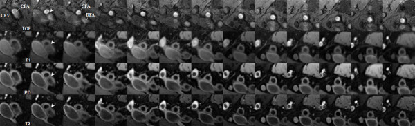
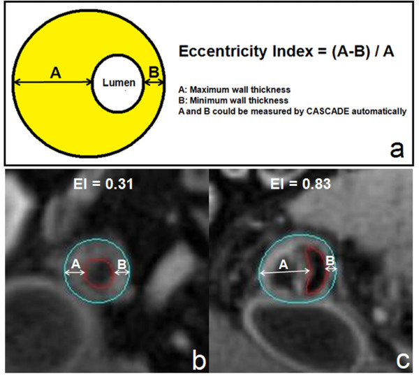
Methods: Twenty-eight subjects with an ankle-brachial index less than 1.00 were examined with 1.5 T high-spatial-resolution, multi-contrast weighted CMR. One hundred and eighty diseased locations of the proximal superficial femoral artery (about 40 mm) were analyzed. The eccentric lesion was defined as [(Maximum wall thickness- Minimum wall thickness)/Maximum wall thickness] >or= 0.5. The arterial morphology and plaque components were measured using semi-automatic image analysis software.
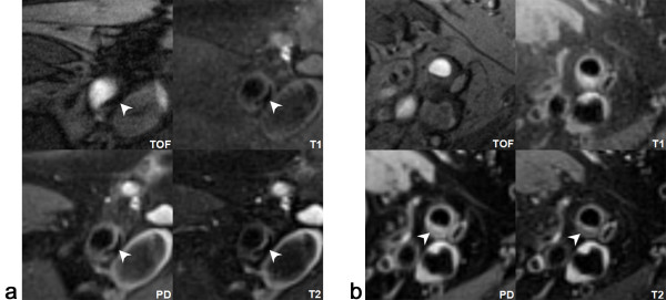
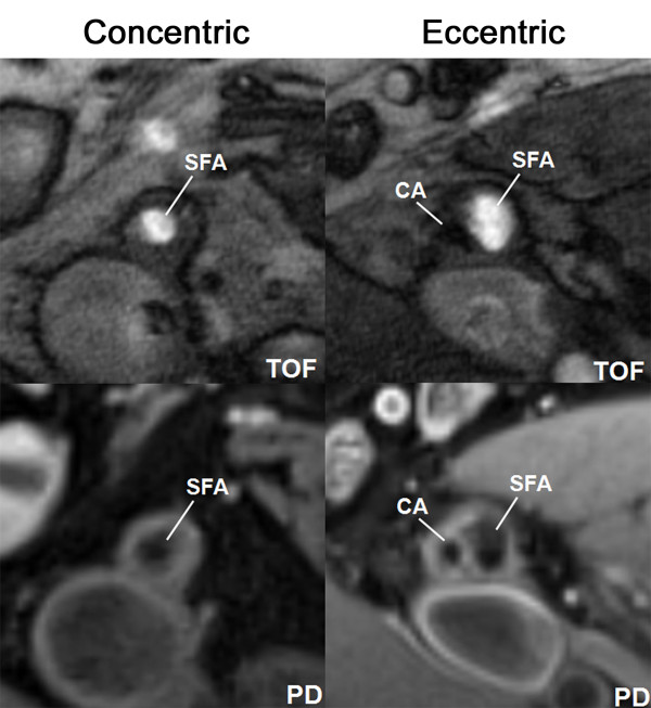
Results: One hundred and fifteen locations were identified as eccentric lesions and sixty-five as concentric lesions. The eccentric lesions had larger wall but similar lumen areas, larger mean and maximum wall thicknesses, and more calcification and lipid rich necrotic core, compared to concentric lesions. For lesions with the same lumen area, the degree of eccentricity was associated with an increased wall area. Eccentricity (dichotomous as eccentric or concentric) was independently correlated with the prevalence of calcification (odds ratio 3.78, 95% CI 1.47-9.70) after adjustment for atherosclerotic risk factors and wall area.
Conclusions: Plaque eccentricity is associated with preserved lumen size and advanced plaque features such as larger plaque burden, more lipid content, and increased calcification in the superficial femoral artery.
Figures
Similar articles
-
Assessment of longitudinal distribution of subclinical atherosclerosis in femoral arteries by three-dimensional cardiovascular magnetic resonance vessel wall imaging.J Cardiovasc Magn Reson. 2018 Sep 3;20(1):60. doi: 10.1186/s12968-018-0482-7. J Cardiovasc Magn Reson. 2018. PMID: 30173671 Free PMC article.
-
Intra-individual comparison of carotid and femoral atherosclerotic plaque features with in vivo MR plaque imaging.Int J Cardiovasc Imaging. 2015 Dec;31(8):1611-8. doi: 10.1007/s10554-015-0737-4. Epub 2015 Aug 22. Int J Cardiovasc Imaging. 2015. PMID: 26296806
-
In Vitro Assessment of Histology Verified Intracranial Atherosclerotic Disease by 1.5T Magnetic Resonance Imaging: Concentric or Eccentric?Stroke. 2016 Feb;47(2):527-30. doi: 10.1161/STROKEAHA.115.011086. Epub 2015 Dec 1. Stroke. 2016. PMID: 26628387
-
Reproducibility of carotid atherosclerotic lesion type characterization using high resolution multicontrast weighted cardiovascular magnetic resonance.J Cardiovasc Magn Reson. 2006;8(6):793-9. doi: 10.1080/10976640600777587. J Cardiovasc Magn Reson. 2006. PMID: 17060101
-
Diabetes-specific characteristics of atherosclerotic plaques in femoral arteries determined by three-dimensional magnetic resonance vessel wall imaging.Diabetes Metab Res Rev. 2020 Jan;36(1):e3201. doi: 10.1002/dmrr.3201. Epub 2019 Jul 24. Diabetes Metab Res Rev. 2020. PMID: 31278827
Cited by
-
In vitro shear stress measurements using particle image velocimetry in a family of carotid artery models: effect of stenosis severity, plaque eccentricity, and ulceration.PLoS One. 2014 Jul 9;9(7):e98209. doi: 10.1371/journal.pone.0098209. eCollection 2014. PLoS One. 2014. PMID: 25007248 Free PMC article.
-
Magnetic Resonance Imaging Techniques in Peripheral Arterial Disease.Adv Wound Care (New Rochelle). 2023 Nov;12(11):611-625. doi: 10.1089/wound.2022.0161. Epub 2023 May 23. Adv Wound Care (New Rochelle). 2023. PMID: 37058352 Free PMC article. Review.
-
Local coronary wall eccentricity and endothelial function are closely related in patients with atherosclerotic coronary artery disease.J Cardiovasc Magn Reson. 2017 Jul 6;19(1):51. doi: 10.1186/s12968-017-0358-2. J Cardiovasc Magn Reson. 2017. PMID: 28679397 Free PMC article.
-
Morphologic characteristics of atherosclerotic middle cerebral arteries on 3T high-resolution MRI.AJNR Am J Neuroradiol. 2013 Sep;34(9):1717-22. doi: 10.3174/ajnr.A3573. Epub 2013 May 2. AJNR Am J Neuroradiol. 2013. PMID: 23639560 Free PMC article.
-
Recent advances in magnetic resonance imaging for peripheral artery disease.Vasc Med. 2018 Apr;23(2):143-152. doi: 10.1177/1358863X18754694. Vasc Med. 2018. PMID: 29633922 Free PMC article. Review.
References
-
- Hirsch AT, Criqui MH, Treat-Jacobson D, Regensteiner JG, Creager MA, Olin JW, Krook SH, Hunninghake DB, Comerota AJ, Walsh ME, McDermott MM, Hiatt WR. Peripheral arterial disease detection, awareness, and treatment in primary care. JAMA. 2001;286:1317–1324. doi: 10.1001/jama.286.11.1317. - DOI - PubMed
-
- Criqui MH, Langer RD, Fronek A, Feigelson HS, Klauber MR, McCann TJ, Browner D. Mortality over a period of 10 years in patients with peripheral arterial disease. N Engl J Med. 1992;326:381–386. - PubMed
-
- Takaya N, Yuan C, Chu B, Saam T, Underhill H, Cai J, Tran N, Polissar NL, Isaac C, Ferguson MS, Garden GA, Cramer SC, Maravilla KR, Hashimoto B, Hatsukami TS. Association between carotid plaque characteristics and subsequent ischemic cerebrovascular events: a prospective assessment with MRI--initial results. Stroke. 2006;37:818–823. doi: 10.1161/01.STR.0000204638.91099.91. - DOI - PubMed
Publication types
MeSH terms
Grants and funding
LinkOut - more resources
Full Text Sources
Medical
