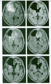
Intra-arterial Chemotherapy for Malignant Tumors of Head and Neck Region Using Three Types of Modified Injection Method
- PMID: 20591239
- PMCID: PMC3553465
- DOI: 10.1177/15910199030090S115
Intra-arterial Chemotherapy for Malignant Tumors of Head and Neck Region Using Three Types of Modified Injection Method
Abstract
Relatively higher infusion rate in the intra-arterial chemotherapy (IA chemotherapy) could induce the higher concentration and the more sufficient distribution of chemotherapeutic agents on tumors. To get the relatively higher infusion rate in IA chemotherapy, we used three types of injection method: high-flow injection, high-dose injection with detoxification and flowcontrolled injection method for the treatment of malignant brain tumors, skull base tumors and head and neck tumors. Between January 1997 and October 2001, twenty-seven patients (mean age 61 y.o.) with supratentorial glioblastoma (4 cases), supratentorial anaplastic astrocytoma (1), CNS lymphoma (2), matastatic skull base tumors (3), and neck tumors (15 squamous cell carcinoma, 1 malignant melanoma and 1 neuroblastoma) received our three types of IA chemotherapy. Sixty- five consecutive procedures were performed. Conventional radiation therapy and/or surgical removal were performed in some of these patients. The median follow-up period was 10 months ranging 2 to 56 months. Fifteen (55.6%) and 6 (22.2%) of 27 patients achieved complete response (CR) and partial response (PR) respectively after initial treatment [CR+PR: 21 (77.8%)]. All responded patients showed clinical improvement. The response rate declined to 55.6% at the end of follow-up period. Eighteen patients are still alive and 15 of them show no evidence of local recurrence. The median post treatment survival was 12 months. There was no serious complication except transient nausea in 4 of 27 (14%) patients, vertigo and granulocytopenia in 1 each (3%) of 27 patients. Our modified IA chemotherapy has provided favorable clinical and radiological results without technical difficulties and serious complications.
Figures


Similar articles
-
Intra-arterial high-dose chemotherapy with cisplatin as part of a palliative treatment concept in oral cancer.AJNR Am J Neuroradiol. 2005 Aug;26(7):1804-9. AJNR Am J Neuroradiol. 2005. PMID: 16091533 Free PMC article. Clinical Trial.
-
High-dose intra-arterial cisplatin therapy followed by radiation therapy for advanced squamous cell carcinoma of the head and neck.Arch Otolaryngol Head Neck Surg. 2001 Jul;127(7):809-12. Arch Otolaryngol Head Neck Surg. 2001. PMID: 11448355
-
Phase II study of induction chemotherapy with paclitaxel, ifosfamide, and carboplatin (TIC) for patients with locally advanced squamous cell carcinoma of the head and neck.Cancer. 2002 Jul 15;95(2):322-30. doi: 10.1002/cncr.10661. Cancer. 2002. PMID: 12124833 Clinical Trial.
-
INGN 201: Ad-p53, Ad5CMV-p53, adenoviral p53, p53 gene therapy--introgen, RPR/INGN 201.Drugs R D. 2007;8(3):176-87. doi: 10.2165/00126839-200708030-00005. Drugs R D. 2007. PMID: 17472413 Review.
-
The application of sentinel node radiolocalization to solid tumors of the head and neck: a 10-year experience.Laryngoscope. 2004 Jan;114(1):2-19. doi: 10.1097/00005537-200401000-00002. Laryngoscope. 2004. PMID: 14709988 Review.
Cited by
-
Current innovations in head and neck cancer: From diagnostics to therapeutics.Oncol Res. 2025 Apr 18;33(5):1019-1032. doi: 10.32604/or.2025.060601. eCollection 2025. Oncol Res. 2025. PMID: 40296914 Free PMC article. Review.
References
-
- Aoki S, Terada H, et al. Supraophthalmic chemotherapy with long tapered catheter: distribution evaluated with intraarterial and intravenous Tc-99m HMPAO. Radiology. 1993;188(2):347–350. - PubMed
-
- Diaz EMJr, Kies MS. Chemotherapy for skull base cancers. Otolaryngol Clin North Am. 2001;34(6):1079–1085. - PubMed
-
- Doolittle ND, Miner ME, et al. Safety and efficacy of a multicenter study using intraarterial chemotherapy in conjunction with osmotic opening of the blood-brain barrier for the treatment of patients with malignant brain tumors. Cancer. 2000;88(3):637–647. - PubMed
-
- Fine HA, Dear KB, et al. Meta-analysis of radiation therapy with and without adjuvant chemotherapy for malignant gliomas in adults. Cancer. 1993;71:2585–2597. - PubMed
-
- Gelman M, Chakeres D, et al. Brain tumors: complications of cerebral angiography accompanied by intraarterial chemotherapy. Neuroradiology. 1999;213:135–140. - PubMed
LinkOut - more resources
Full Text Sources
Research Materials

