

Transarterial Wedged-catheter, Flow-arrest, N-butyl Cyanoacrylate Embolization of Three Dural Arteriovenous Fistulae in a Single Patient
- PMID: 20591254
- PMCID: PMC3548213
- DOI: 10.1177/159101990300900307
Transarterial Wedged-catheter, Flow-arrest, N-butyl Cyanoacrylate Embolization of Three Dural Arteriovenous Fistulae in a Single Patient
Abstract
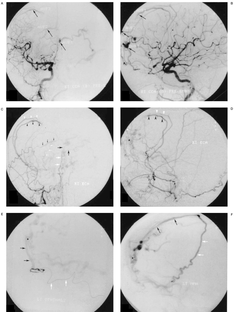
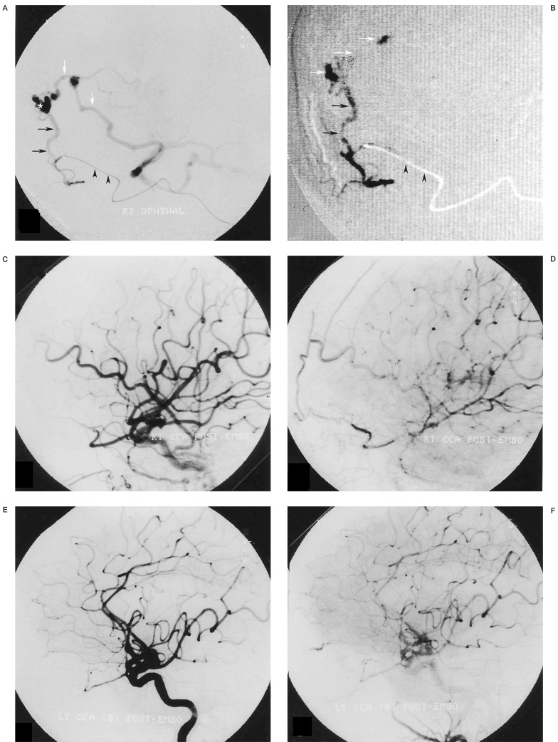
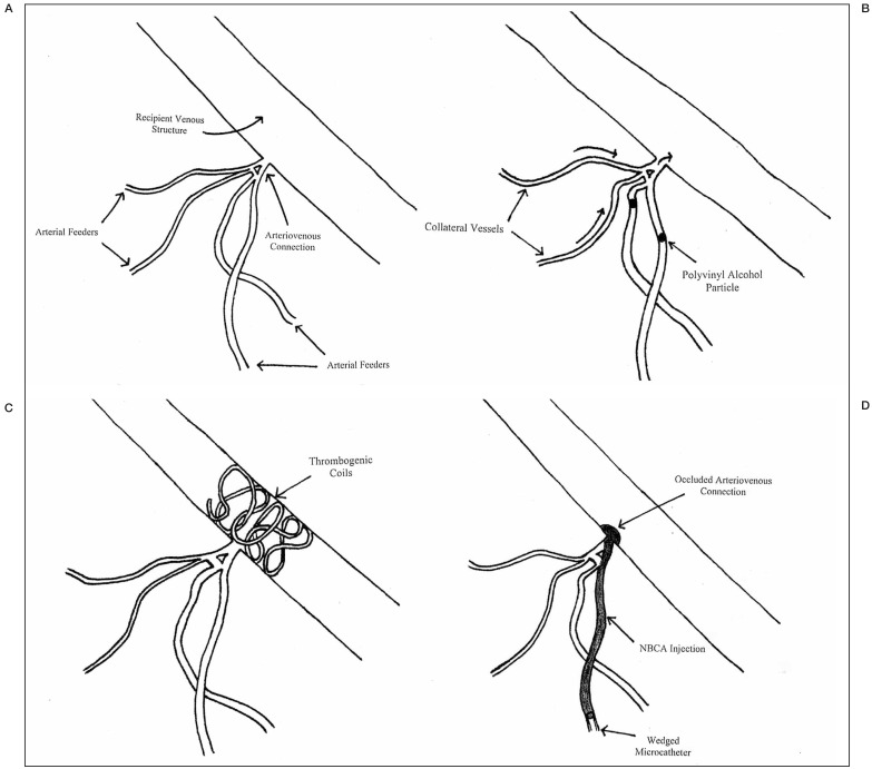
The pathogenesis of dural arteriovenous fistulas (DAVFs) is currently unknown, with multiple DAVFs being rare. For patients with limited venous access secondary to sinus thrombosis, or for patients where parent sinus occlusion would not be tolerated, transvenous embolization may not be possible and other treatment methods must be considered. A 69-year-old female patient with a two-year history of progressive headaches, memory loss, and unsteady gait underwent cerebral angiography that revealed three separate DAVFs with congested cortical venous drainage overlying both frontal lobes. Using an application of a transarterial wedged-catheter, flow-arrest technique, N-butyl cyanoacrylate was deposited across all three pathologic arteriovenous connections providing a definitive cure. Transarterial NBCA embolization may provide curative treatment of DAVFs, and is of particular utility in situations where access to the draining venous structures is limited.
Figures
Similar articles
-
Use of a wedged microcatheter for curative transarterial embolization of complex intracranial dural arteriovenous fistulas: indications, endovascular technique, and outcome in 21 patients.J Neurosurg. 2003 Mar;98(3):498-506. doi: 10.3171/jns.2003.98.3.0498. J Neurosurg. 2003. PMID: 12650420
-
Dural arteriovenous fistulas: a review of the literature and a presentation of a single institution's experience.World Neurosurg. 2013 Jul-Aug;80(1-2):94-102. doi: 10.1016/j.wneu.2012.01.053. Epub 2012 Jan 31. World Neurosurg. 2013. PMID: 22381858
-
Transarterial embolization of clival dural arteriovenous fistulae using liquid embolic agents.Neurosurgery. 2008 Feb;62(2):408-15; discussion 415. doi: 10.1227/01.neu.0000316007.34259.26. Neurosurgery. 2008. PMID: 18382318
-
Multiple Dural and Pial Arteriovenous Fistulae in a Twenty-Four-Year-Old Woman in the Setting of Superior Sagittal Sinus Thrombosis: Case Report and Review of Literature.J Stroke Cerebrovasc Dis. 2016 Oct;25(10):e192-9. doi: 10.1016/j.jstrokecerebrovasdis.2016.07.037. Epub 2016 Aug 17. J Stroke Cerebrovasc Dis. 2016. PMID: 27544867 Review.
-
Endovascular treatment of the cavernous sinus dural arteriovenous fistula: current status and considerations.Int J Med Sci. 2020 May 1;17(8):1121-1130. doi: 10.7150/ijms.45210. eCollection 2020. Int J Med Sci. 2020. PMID: 32410842 Free PMC article. Review.
Cited by
-
Combined transarterial and transvenous embolization of anterior cranial fossa dural arteriovenous fistula.Surg Neurol Int. 2023 Aug 4;14:277. doi: 10.25259/SNI_487_2023. eCollection 2023. Surg Neurol Int. 2023. PMID: 37680916 Free PMC article.
-
Regression of a Flow-Related Ophthalmic Artery Aneurysm After Treatment of a Frontal DAVS. A Case Report.Interv Neuroradiol. 2004 Sep 30;10(3):265-8. doi: 10.1177/159101990401000310. Epub 2005 Jan 5. Interv Neuroradiol. 2004. PMID: 20587240 Free PMC article.
-
Progress in research on intracranial multiple dural arteriovenous fistulas.Biomed Rep. 2018 Jan;8(1):17-25. doi: 10.3892/br.2017.1021. Epub 2017 Nov 21. Biomed Rep. 2018. PMID: 29399335 Free PMC article.
References
-
- Cognard C, Gobin Y, et al. Cerebral dural arteriovenous fistulas: clinical and angiographic correlation with a revised classification of venous drainage. Radiology. 1995;194:671–680. - PubMed
-
- Chaloupka J, Huddle D, Alderman J. Local induction of angiogenesis within the walls of a dural sinus results in the creation of dural arteriovenous fistula: demonstration by a polymeric release implantation model in swine (abstract) J Neurosurg. 1999;90:194A.
-
- Chaloupka J, Marx W, Kallmes D. Dural arteriovenous fistulas. J Neurosurg. 2001;94:858–860. - PubMed
-
- Folkman J, Klagsbrun M. Angiogenic factors. Science. 1987;235:442–447. - PubMed
LinkOut - more resources
Full Text Sources
