

doi: 10.1177/159101990300900209.
Epub 2004 Oct 22.
Two Distally Located Right SCA Aneurysms: Endovascular Treatment by Parent Artery Occlusion with GDC Coils and N-BCA Injection. Case Report and Review of the Literature
Affiliations
- PMID: 20591270
- PMCID: PMC3547514
- DOI: 10.1177/159101990300900209
Item in Clipboard
Two Distally Located Right SCA Aneurysms: Endovascular Treatment by Parent Artery Occlusion with GDC Coils and N-BCA Injection. Case Report and Review of the Literature
Interv Neuroradiol.
.
Abstract
Peripheral aneurysms of the superior cerebellar artery are considered difficult to treat surgically and endovascularly because of their inaccessibility. Parent artery occlusion is therefore frequently the preferred method. Embolic materials previously reported in this situation are either GDC coils or a polymerizing agent (n- BCA). We report a patient with two distally located, wide-neck aneurysms of the right superior cerebellar artery who presented with hemorrhage and was treated by endovascular embolization of the parent artery using a combination of GDC coils and n-BCA.
Figures

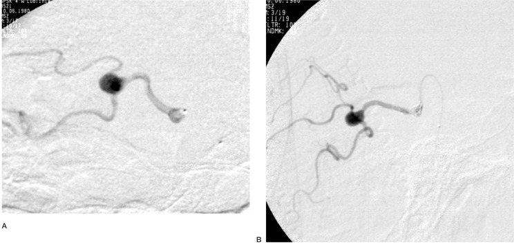
Left vertebral artery injection, A-P (A) and oblique (B) projections showing two aneurysms on the inferior branch of left superior cerebellar artery.
Selective angiogram - working projection before coil embolization and glue injection. Microcatheter tip placed in the sac of the aneurysm (A). Selective angiogram performed after two GDC 10 coils have been delivered shows that the parent artery is still open (B). At this point, the decision to use glue injection was made and the tip of microcatheter slightly withdrawn to gain better flow conditions.

Lateral X ray view after treatment,showing GDC 10 coils in the sac of the proximal aneurysm, cast of glue in the parent artery and partially in the sac of the distal aneurysm where the stasis of the contrast is seen.

A-B Post-treatment control angiograms of the left vertebral artery: A-P (A) and oblique (B) showing complete selective occlusion of the inferior branch of left superior cerebellar artery as well as two aneurysms.
Similar articles
-
Endovascular treatment of peripheral intracranial aneurysms.AJNR Am J Neuroradiol. 2007 Feb;28(2):355-61. AJNR Am J Neuroradiol. 2007. PMID: 17297012 Free PMC article.
-
Parent artery occlusion for intracranial aneurysms.Interv Neuroradiol. 2009 Sep;15(3):309-15. doi: 10.1177/159101990901500308. Epub 2009 Nov 4. Interv Neuroradiol. 2009. PMID: 20465914 Free PMC article.
-
Endovascular management of distal anterior inferior cerebellar artery aneurysms: Report of two cases and review of the literature.Surg Neurol Int. 2011;2:95. doi: 10.4103/2152-7806.82577. Epub 2011 Jun 30. Surg Neurol Int. 2011. PMID: 21748047 Free PMC article.
-
Aneurysm arising from the cortical segment of the superior cerebellar artery: a case report and review of the literatures.Surg Neurol. 2008 Oct;70(4):421-4; discussion 424. doi: 10.1016/j.surneu.2007.02.061. Epub 2008 Mar 4. Surg Neurol. 2008. PMID: 18291446 Review.
-
Endovascular Treatment for Peripheral Superior Cerebellar Artery Aneurysms: Current State and Future Considerations.World Neurosurg. 2019 Jul;127:423-433. doi: 10.1016/j.wneu.2019.04.145. Epub 2019 Apr 25. World Neurosurg. 2019. PMID: 31028980 Review.
References
-
- Yoshimoto T, Kayama T, et al. Distribution of intracranial aneurysm. In: Suzuki J, editor. Cerebral Aneurysms Neuron. Tokyo, Japan: 1979. pp. 14–19.
-
- Locksley HB. Report on the cooperative study of intracranial aneurysms and subarachnoid hemorrhage, section V, part 1: natural history of subarachnoid hemorrhage, intracranial aneurysms and arteriovenous malformations: based on 6368 cases in the cooperative study. J Neurosurg. 1983;58:350–352. - PubMed
LinkOut - more resources
Full Text Sources
